

Experimental research of monochromatic X-ray microscopy
-
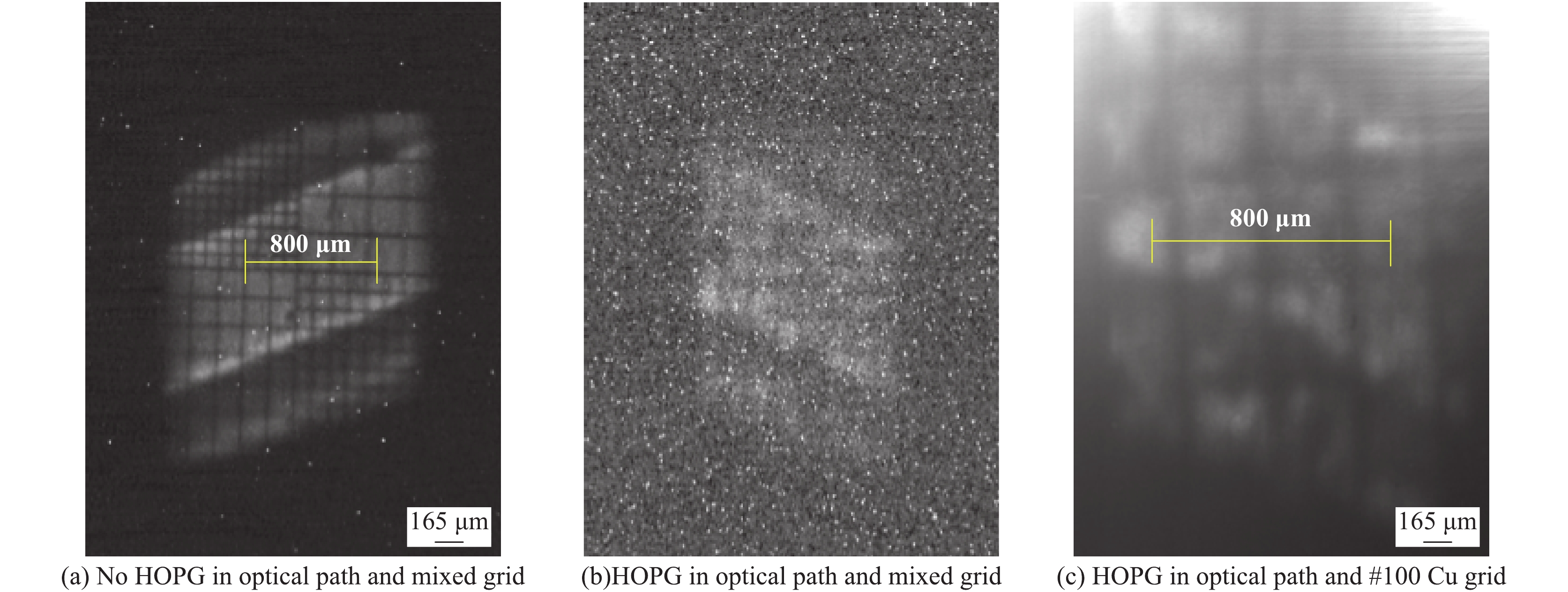
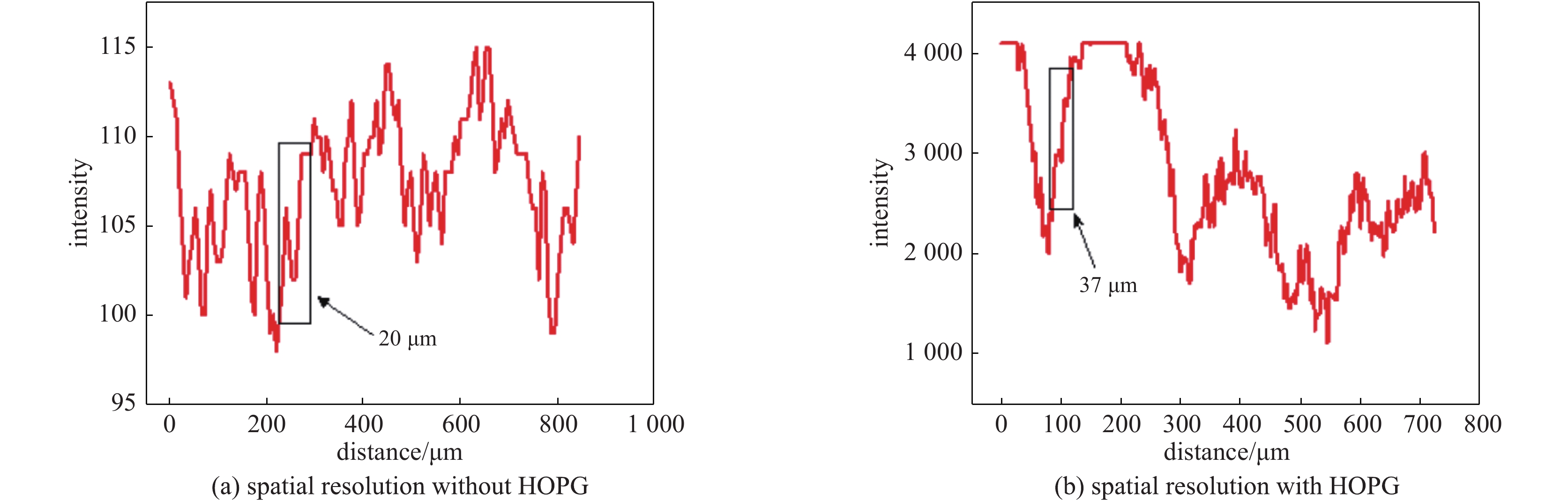
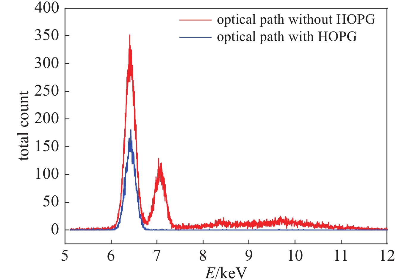
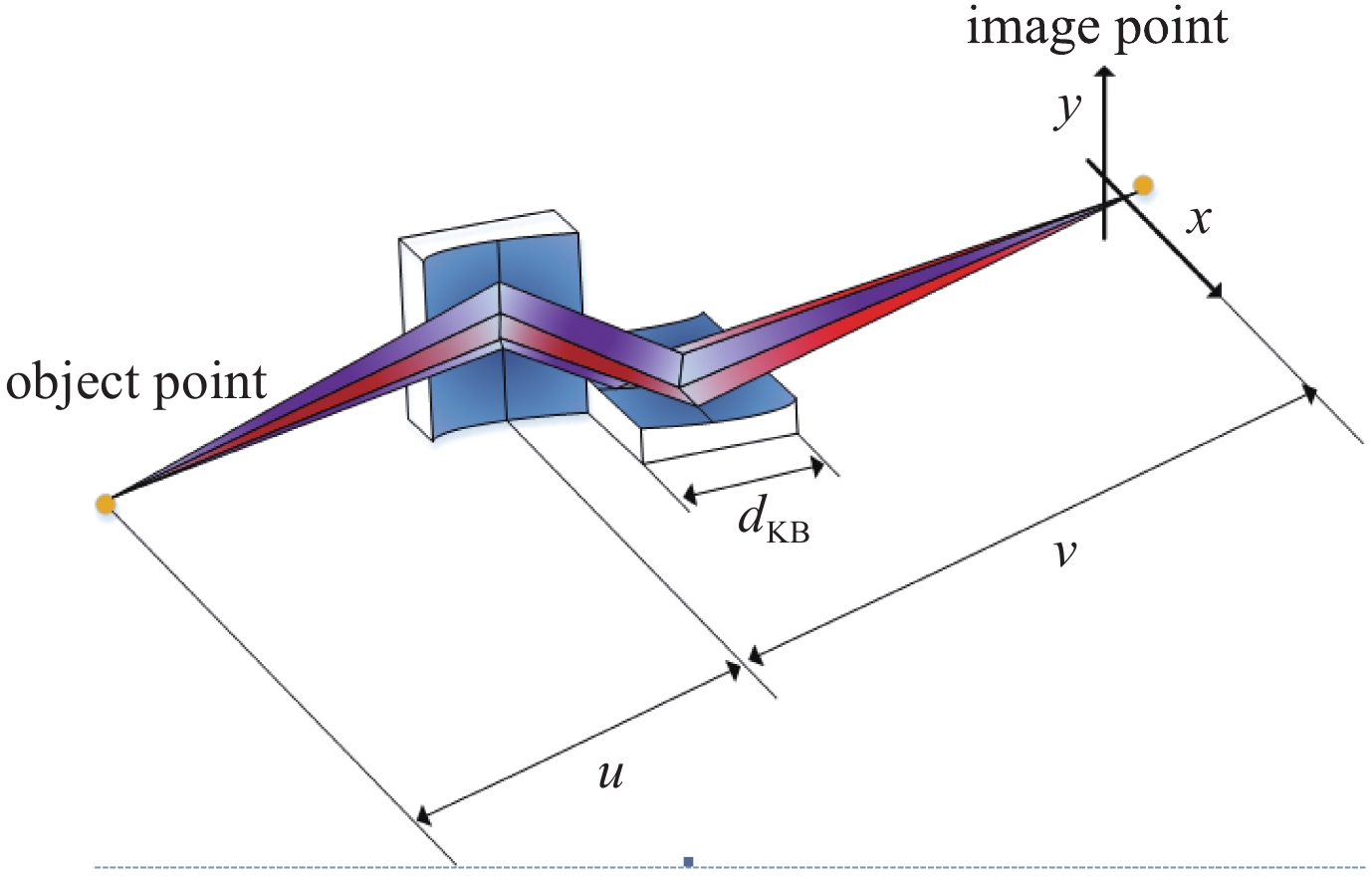
摘要: 围绕激光惯性约束聚变(ICF)内爆压缩阶段高空间分辨、高能谱分辨的诊断需求,提出了一种将KB显微镜和衍射晶体组合的大视场、单色化成像系统。在实验室条件下,利用Fe靶X射线光管,采用KB显微镜结合高定向热解石墨(HOPG)对网格进行背光成像,晶体选能后的成像结果表明,系统的视场能达到800 μm,其中高分辨区域成像的分辨率为37 μm。采用能谱探测器测试成像能谱,结果表明,系统的能量分辨率为28,验证了系统的单色性能。该系统兼顾了大视场、空间分辨和能量分辨,对内爆压缩阶段实验中热斑结构及混合效应的研究具有重要应用。Abstract: Based on the diagnostic requirements of high spatial resolution and high energy spectral resolution in the implosion compression stage of laser inertial confinement fusion (ICF), this paper proposes a large field of view and monochromatic imaging system, which combines KB microscope and diffractive crystal. Under the laboratory condition, the grid is backlight imaged by using the Fe target X-ray tube, with KB microscope and high oriented pyrolytic graphite (HOPG). The imaging results after the crystal energy selection show that the field of view of the system can reach 800 μm, and the resolution of high-resolution area imaging is 37 μm. The detecting results show that the energy resolution of the system is 28, which verifies the monochromatic performance of the system. The system takes into account large field of view, spatial resolution and energy resolution, and has an important application in the research of hot spot structure and mixing effect in the experiment of implosion compression stage.
-
Key words:
- KB microsope /
- diffractive crystal /
- monochromatic imaging /
- energy spectrum resolution
-
表 1 KB结构参数
Table 1. Structure parameters of KB microscope
No. E/ keV R/m u/mm v/mm M θ/(°) mirror1 6.4 50.0 200 800 4.0 0.43 mirror2 6.4 50.0 210 790 3.8 0.42  下载: 导出CSV
-
[1] Lindl J D, Amendt P, Berger R L, et al. The physics basis for ignition using indirect-drive targets on the National Ignition Facility[J]. Physics of Plasmas, 2004, 11(2): 339-491. doi: 10.1063/1.1578638 [2] 蒲昱东, 陈伯伦, 黄天晅, 等. 激光间接驱动惯性约束聚变内爆物理实验研究[J]. 强激光与粒子束, 2015, 27:032015. (Pu Yudong, Chen Bolun, Huang Tianxuan, et al. Experimental studies of implosion physics of indirect drive inertial confinement fusion[J]. High Power Laser and Particle Beams, 2015, 27: 032015 doi: 10.11884/HPLPB201527.032015 [3] 陈伯伦, 韦敏习, 杨正华, 等. 球面弯晶的背光成像特性[J]. 强激光与粒子束, 2013, 25(3):641-645. (Chen Bolun, Wei Minxi, Yang Zhenghua, et al. Character of backlight imaging based on spherically bent crystal[J]. High Power Laser and Particle Beams, 2013, 25(3): 641-645 doi: 10.3788/HPLPB20132503.0641 [4] 张强强, 魏来, 杨祖华, 等. 用于超热电子诊断的单色X射线成像技术[J]. 光学学报, 2016, 36:1234001. (Zhang Qiangqiang, Wei Lai, Yang Zuhua, et al. Monochromatic X-ray imaging technology for diagnostics of hot electrons[J]. Acta Optica Sinica, 2016, 36: 1234001 [5] 忻秋琪, 李亚冉, 陈亮, 等. 四通道球面弯晶成像系统设计及实验研究[J]. 强激光与粒子束, 2019, 31:052001. (Qi Qiuqi, Li Yaran, Chen Liang, et al. Design and experimental research of four-channel spherically bent crystal imaging system[J]. High Power Laser and Particle Beams, 2019, 31: 052001 doi: 10.11884/HPLPB201931.190006 [6] Christensen F E. X-ray multilayers in diffractometers, monochromators, and spectrometers[M]. Technical Symposium. 1988: 124-132. [7] Jiang H. Design and characterization of EUV and X-ray multilayers[M]//Optimization Algorithms-Methods and Applications. 2016. [8] Li Y, Mu B, Xie Q, et al. Development of an x-ray eight-image Kirkpatrick-Baez diagnostic system for China’s laser fusion facility[J]. Applied optics, 2017, 56(12): 3311-3318. doi: 10.1364/AO.56.003311 [9] Koch J A, Landen O L, Barbee T W, et al. High-energy X-ray microscopy techniques for laser-fusion plasma research at the National Ignition Facility[J]. Applied Optics, 1998, 37(10): 1784-1795. doi: 10.1364/AO.37.001784 [10] 蔡厚智, 刘进元, 彭翔, 等. 宽微带X射线分幅相机的研制[J]. 中国激光, 2012, 39(1):241-247. (Cai Houzhi, Liu Jinyuan, Peng Xiang, et al. Development of wide microstrip X-ray framing camera[J]. China Laser, 2012, 39(1): 241-247 [11] 单宝忠, 郭宝平, 牛憨笨. 多通道门选通纳秒分幅相机[J]. 光学精密工程, 2007, 15(12):1963-1968. (Shan Baozhong, Guo Baoping, Niu Hanben. Multi pass gate gated nanosecond framing camera[J]. Optical Precision Engineering, 2007, 15(12): 1963-1968 doi: 10.3321/j.issn:1004-924x.2007.12.022 [12] 成金秀, 常增虎. MCP选通X射线皮秒分幅相机研制进展[J]. 光学精密工程, 1996, 4(1):44-48. (Cheng Jinxiu, Chang Zenghu. Development of MCP gated X-ray picosecond framing camera[J]. Optical Precision Engineering, 1996, 4(1): 44-48 doi: 10.3321/j.issn:1004-924X.1996.01.010 [13] Marshall F J, Su Q. Quantitative measurements with X-ray microscopes in laser-fusion experiments[J]. Review of Scientific Instruments, 1995, 66(1): 725. doi: 10.1063/1.1146270 [14] Marshall F J, Oertel J A. A framed monochromatic X-ray microscope for ICF (invited)[J]. Review of Scientific Instruments, 1997, 68(1): 735. doi: 10.1063/1.1147688 [15] Marshall F J, Delettrez J A, Meyerhofer D D, et al. Monochromatic imaging of direct-drive implosions on OMEGA[C]//APS Meeting. 2000. [16] Xie Q, Mu B, Li Y, et al. Development of high resolution dual-energy KBA microscope with large field of view for RT-instability diagnostics at SG-III facility[J]. Optics Express, 2017, 25(3): 2608. doi: 10.1364/OE.25.002608 -

 点击查看大图
图(9) / 表(1)
计量
- 文章访问数: 1434
- HTML全文浏览量: 384
- PDF下载量: 68
- 被引次数: 0
