Electric field enhancement effect and damage characteristics of nodular defect in 45° high-reflection coating
-
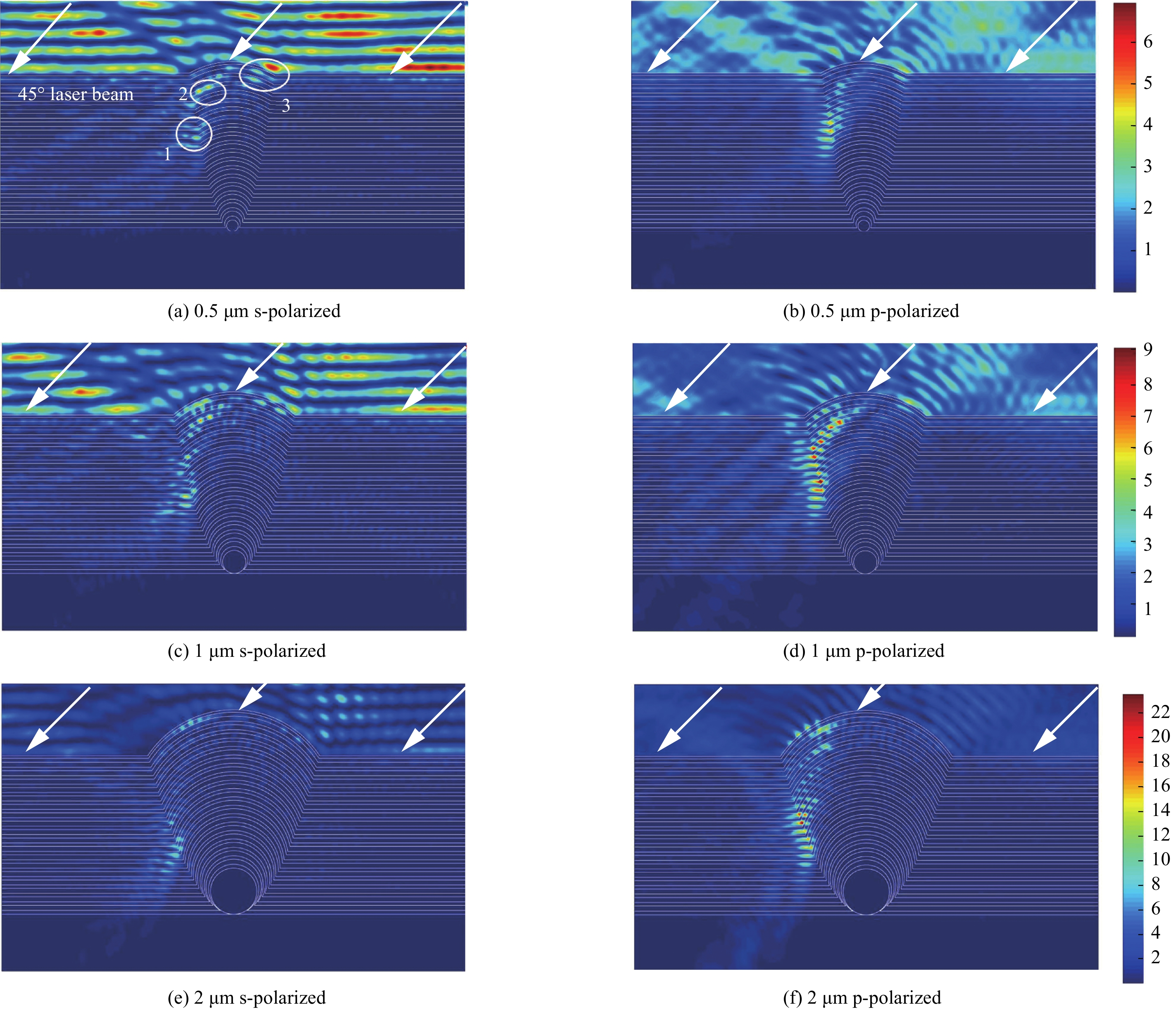
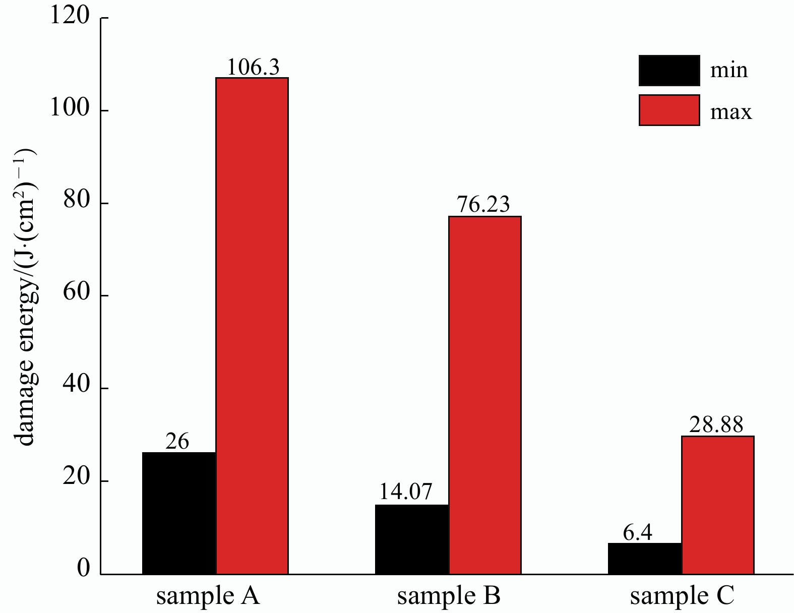
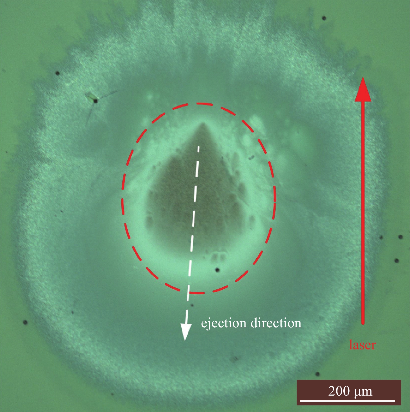
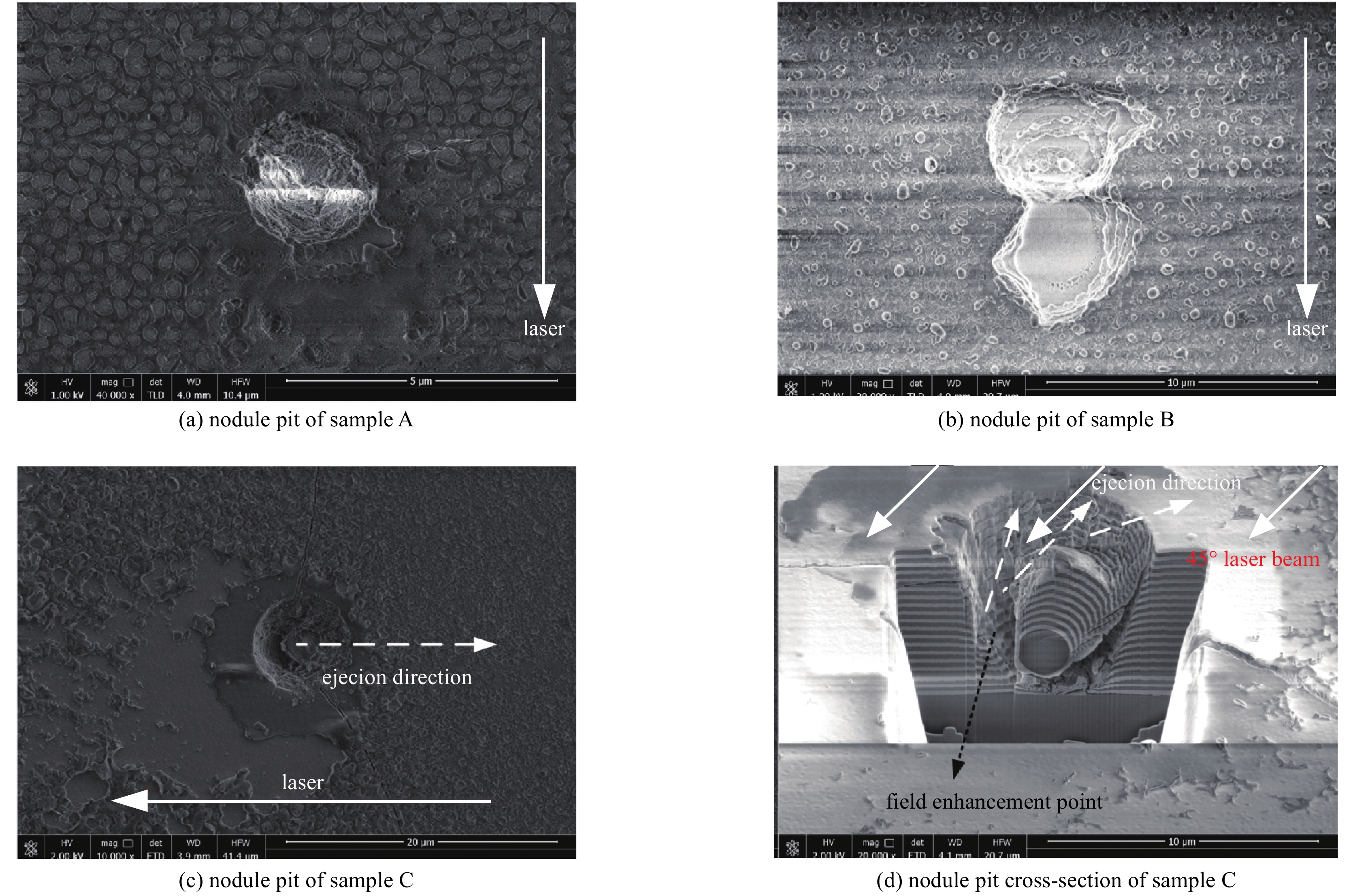
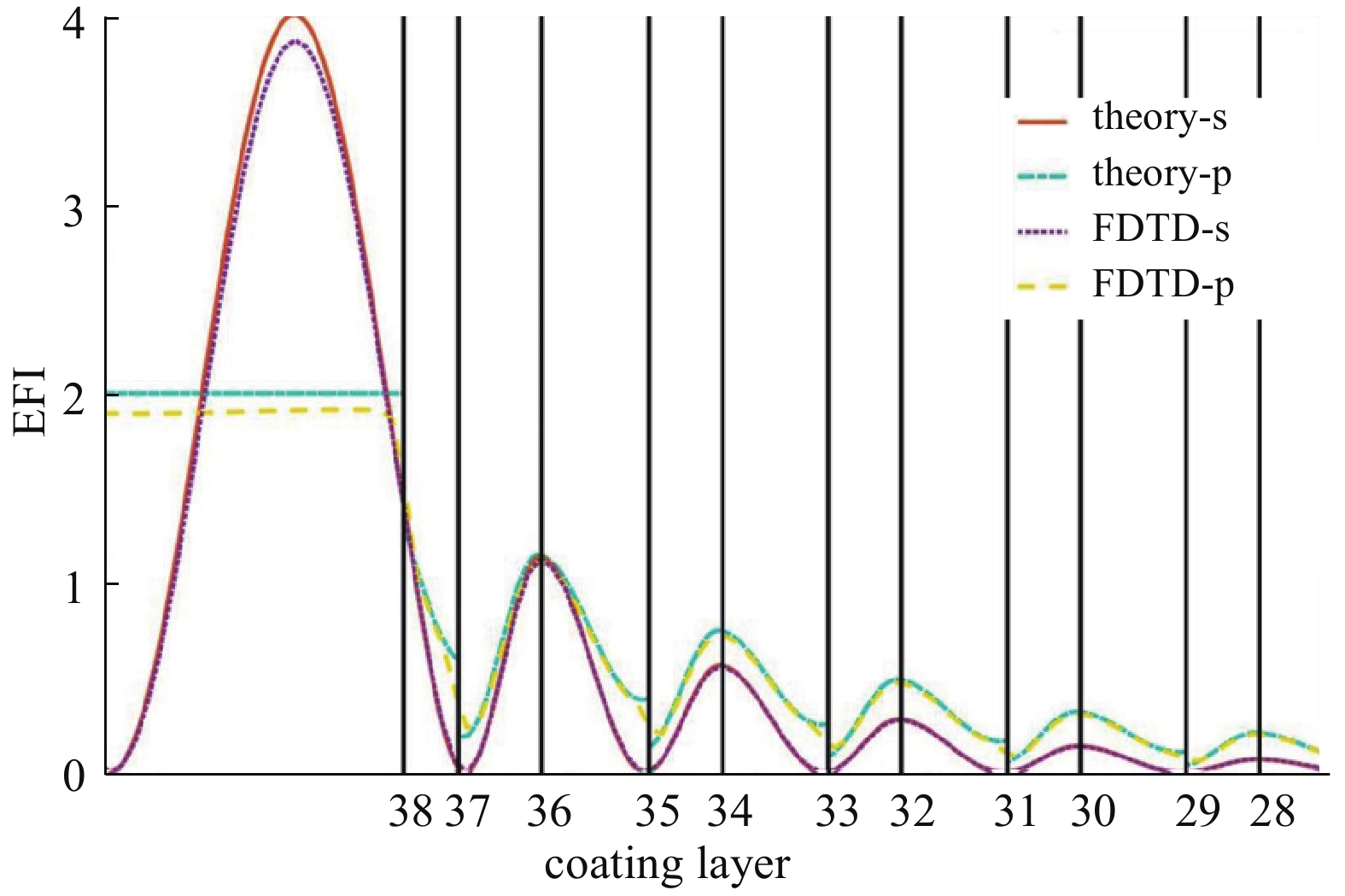
摘要: 研究设计和制备了中心波长为1 064 nm的45°多层膜反射镜,通过数值仿真结合实验,对薄膜中节瘤缺陷引起的电场增强效应及其对薄膜抗激光损伤性能的影响进行了研究。结果表明:当1 064 nm激光从右至左45°斜入射时,电场增强效应主要出现在节瘤缺陷的表层及其左侧轮廓中部,电场增强效应随节瘤缺陷尺寸增大而增强。实验上,在清洁的基板表面喷布单分散SiO2微球作为人工节瘤种子,采用电子束蒸发制备法完成多层全反膜的制备,采用R-on-1方式对薄膜样品进行激光损伤测试。结果表明,薄膜的损伤阈值随着节瘤缺陷尺寸增加而减小。通过综合分析电场增强效应、薄膜损伤测试结果及损伤形貌特征得出,薄膜损伤阈值降低是由于节瘤缺陷和薄膜中微缺陷共同作用的结果。Abstract: Nodular defect is one of the most common defects that affect the laser damage resistance of optical thin films. It has been an important research object in the field of high power laser thin films at home and abroad. 45° multilayer mirrors with a central wavelength of 1 064 nm were designed and fabricated. Through numerical simulations and experiments, the electric field enhancement effect(EFEE) caused by nodular defect and its influence on the laser damage resistance of the film were studied. The results show that when the 1 064 nm laser is incident obliquely from right to left at 45°, the EFEE mainly appears in the surface layer of the nodular defect and the middle of its left profile. The EFEE increases with the size of nodule defect. In the experiment, mono-disperse SiO2 microspheres were sprayed on the surface of clean substrates as the artificial nodule seeds, the multilayer high reflection coatings were prepared by electron beam evaporation, and the laser damage resistance of the samples was tested by R-on-1 method. By comprehensively analyzing the experiment results, it is concluded that the damage threshold reduction of coatings is due to the nodular defects and micro-defects in the coatings, and the damage threshold of coatings decreases with the increase of nodular defect size.
-
表 1 不同区域的电场强度
Table 1. The electric field intensity in different regions
seed diameter/μm electric field intensity s-polarized p-polarized region 1 region 2 region 3 region 1 region 2 region 3 0.5 3.78 4.59 6.37 5.14 3.06 4.06 1 5.99 6.75 5.65 9.14 8.01 5.63 2 10.43 10.69 7.38 23.55 16.07 5.00 表 2 样品参数
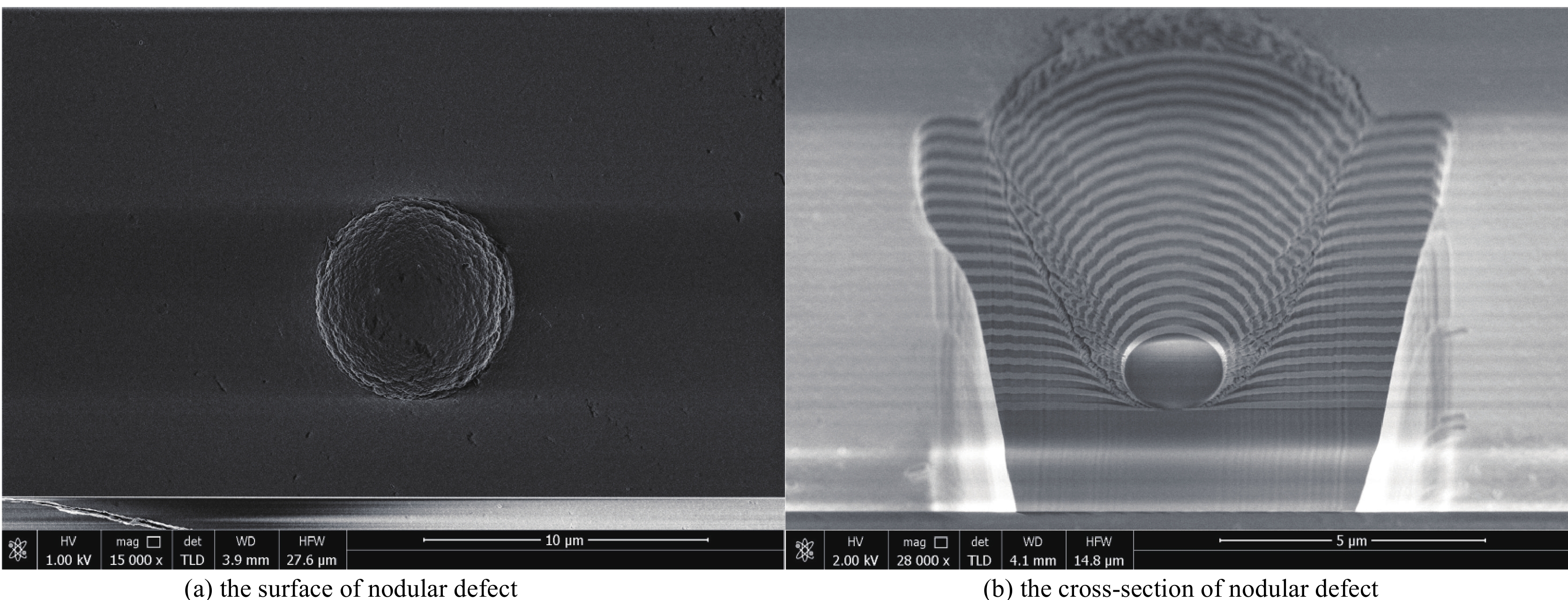
Table 2. Sample parameters
sample number seed diameter d/μm nodule diameter D/μm coating thickness t/μm coefficient c A 0.5 3.5±0.2 ~6.75 3.62 B 1 4.8±0.2 3.41 C 2 7.0±0.2 3.63 -
[1] Koldunov M F, Manenkov A A, Pocotilo I L. Theory of laser-induced damage to optical coatings: inclusion-initiated thermal explosion mechanism[C]//Proc of SPIE. 1994, 2114: 469-487. [2] Kozlowski M R, Chow R. The role of defects in laser damage of multilayer coatings[C]//Proc of SPIE. 1994, 2114: 640-649. [3] Dijon J, Kaiser N, Schallenberg U B, et al. Influence of substrate cleaning on LIDT of 355 nm HR coatings[C]//Proc of SPIE. 1997, 2966: 178-186. [4] Zhang Dawei, Shao Jianda, Fan Shuhai, et al. The effects of ion cleaning on the roughness of substrates and laser induced damage thresholds of films[C]//Optical Interference Coatings. 2004: 345-348 [5] Bevis R P, Sheehan L M, Smith D J, et al. The advantages of evaporation of Hafnium in a reactive environment to manufacture high damage threshold multilayer coatings by electron-beam deposition[C]//Proc of SPIE. 1999, 3738: 318-324. [6] Chow R, Falabella S, Loomis G E, et al. Reactive evapoation of low defect density hafnia[J]. Applied Optics, 1993, 32(28): 5567-5574. doi: 10.1364/AO.32.005567 [7] 谢凌云, 程鑫彬, 张锦龙, 等. 节瘤缺陷激光损伤的研究进展[J]. 强激光与粒子束, 2016, 28:090201. (Xie Lingyun, Cheng Xinbin, Zhang Jinlong, et al. Research process of laser-induced damage of nodular defects[J]. High Power Laser and Particle Beams, 2016, 28: 090201 doi: 10.11884/HPLPB201628.160058 [8] Liu Xiaofeng, Zhao Yuan’an, et al. Characteristics of nodular defect in HfO2/SiO2 multilayer optical coatings[J]. Applied Surface Science, 2010, 256(12): 3783-3788. doi: 10.1016/j.apsusc.2010.01.026 [9] Dijon J, Poulingue M, Hue J. Thermomechanical model of mirror laser damage at 1.06 μm: I. Nodule ejection[C]//Proc of SPIE. 1999, 3578: 387-397. [10] Cheng Xinbin, Wei Zeyong, Zhang Jinlong, et al. Physical insight toward electric field enhancement at nodular defects in optical coatings[J]. Optics Express, 2015, 23(7): 8609-8619. doi: 10.1364/OE.23.008609 [11] Poulingue M, Dijon J, Rafin B, et al. Generation of defects with diamond and silica particles inside high-reflection coatings: influence on the laser damage threshold[C]//Proc of SPIE. 1999, 3738: 325-336. [12] Shan Yongguang, He Hongbo, Wei Chaoyang, et al. Geometrical characteristics and damage morphology of nodules grown from artificial seeds in multilayer coating[J]. Applied Optics, 2010, 49(22): 4290-4295. doi: 10.1364/AO.49.004290 [13] Cheng Xinbin, Lequime M, Macleod H A, et al. Using monodisperse SiO2 microspheres to study laser-induced damage of nodules in HfO2/SiO2 high reflectors[C]//Proc of SPIE. 2011: 816816. [14] Deford J F, Kozlowski M R. Modeling of electric-field enhancement at nodular defects in dielectric mirror coatings[C]//Proc of SPIE. 1993, 1848: 455-472. [15] Cheng Xinbin, Zhang Jinlong, Ding Tao, et al. The effect of an electric field on the thermomechanical damage of nodular defects in dielectric multilayer coatings irradiated by nanosecond laser pulses[J]. Light: Science & Applications, 2013, 2(6): e80. [16] Yee K S. Numerical solution of initial boundary value problems involving Maxwell's equations in isotropic media[J]. IEEE Trans Antennas & Propagation, 1966, 14: 302-307. [17] Hue J, Garrec P, Dijon J, et al. R-on-1 automatic mapping: a new tool for laser damage testing[C]//Proc of SPIE. 1995, 2714: 90-101. [18] Han Jinghua, Li Yaguo, He Changtao, et al. Effects of laser plasma on damage in optical glass induced by pulsed lasers[J]. Optical Engineering, 2012, 51: 121809. doi: 10.1117/1.OE.51.12.121809 [19] 周成虎, 张秋慧, 黄明明, 等. 杂质微粒对薄膜的损伤效应[J]. 红外与激光工程, 2016, 45:0721004. (Zhou Chenghu, Zhang Qiuhui, Huang Mingming, et al. Damage effects of impurity particles on film[J]. Infrared and Laser Engineering, 2016, 45: 0721004 doi: 10.3788/irla201645.0721004 [20] Genin F Y, Stolz C J. Morphologies of laser-induced damage in hafnia-silica multilayer mirror and polarizer coatings[C]//Proc of SPIE. 1996, 2870: 439-448. [21] Zhu Meiping, Yi Kui, Li Dawei, et al. Influence of SiO2 overcoat layer and electric field distribution on laser damage threshold and damage morphology of transport mirror coatings[J]. Optics Communications, 2014, 319: 75-79. doi: 10.1016/j.optcom.2014.01.014 [22] Zhao Yuan’an, Gao Weidong, Shao Jianda, et al. Roles of absorbing defects and structural defects in multilayer under single-shot and multi-shot laser radiation[J]. Applied Surface Science, 2004, 227(1/4): 275-281. [23] Dijon J, Rafin B, Pelle C, et al. One-hundred Joule per square centimeter 1.06-μm mirrors[C]//Proc of SPIE. 2000, 3902: 158-168. [24] Cheng Xinbin, Shen Zhengxiang, Jiao Hongfei, et al. Laser damage study of nodules in electron-beam-evaporated HfO2/SiO2 high reflectors[J]. Applied Optics, 2011, 50(9): 357-363. doi: 10.1364/AO.50.00C357 [25] Liu Xiaofeng, Zhao Yuan’an, Gao Yanqi, et al. Investigations on the catastrophic damage in multilayer dielectric films[J]. Applied Optics, 2013, 52(10): 2194-2199. doi: 10.1364/AO.52.002194 -


 下载:
下载:
