-
Abstract:
The evolution of electromagnetic field and fluid is important in the research of high energy density physics, controlled nuclear fusion, and laboratory astrophysics. But in experiment, it is difficult to get the density and electromagnetic field distribution simultaneously. Based on high energy electron lens radiography, this paper proposes dual degrees of freedom diagnosis (DDFD) by constructing areal density difference. Combining the Monte-Carlo simulation and beam optics analysis, the feasibility of this method when the diagnosed system includes relatively strong electromagnetic field has been validated. Besides, changing the aperture as a ring can effectively improve the resolution in low E/B field and low areal density situation. The simulation results indicate that this method works well. Considering the characters of electron beams, this method is quite suitable for the electromagnetic fluid diagnosis.
摘要:电磁场及流体演化过程信息的获取在高能量密度物理、可控核聚变及实验天体物理的研究中起着重要的作用,然而在实验过程中,电磁场信息及流体信息的同步获取是非常困难的。基于高能电子透镜成像技术,利用面密度差分的方法,提出了一种可以实现面密度和场积分强度同时获取的双自由度诊断设计方案。结合了蒙特卡罗模拟和束流光学分析,该方案在相对较强的电磁场情况下的适用性得到了验证。此外,我们可以通过改变Fourier面处光阑的形状将其适用区间扩展到低电磁场强度相空间。结合高能电子束相对论速度及超短脉冲的特点,该技术非常适用于磁流体超快演化过程的诊断。
-
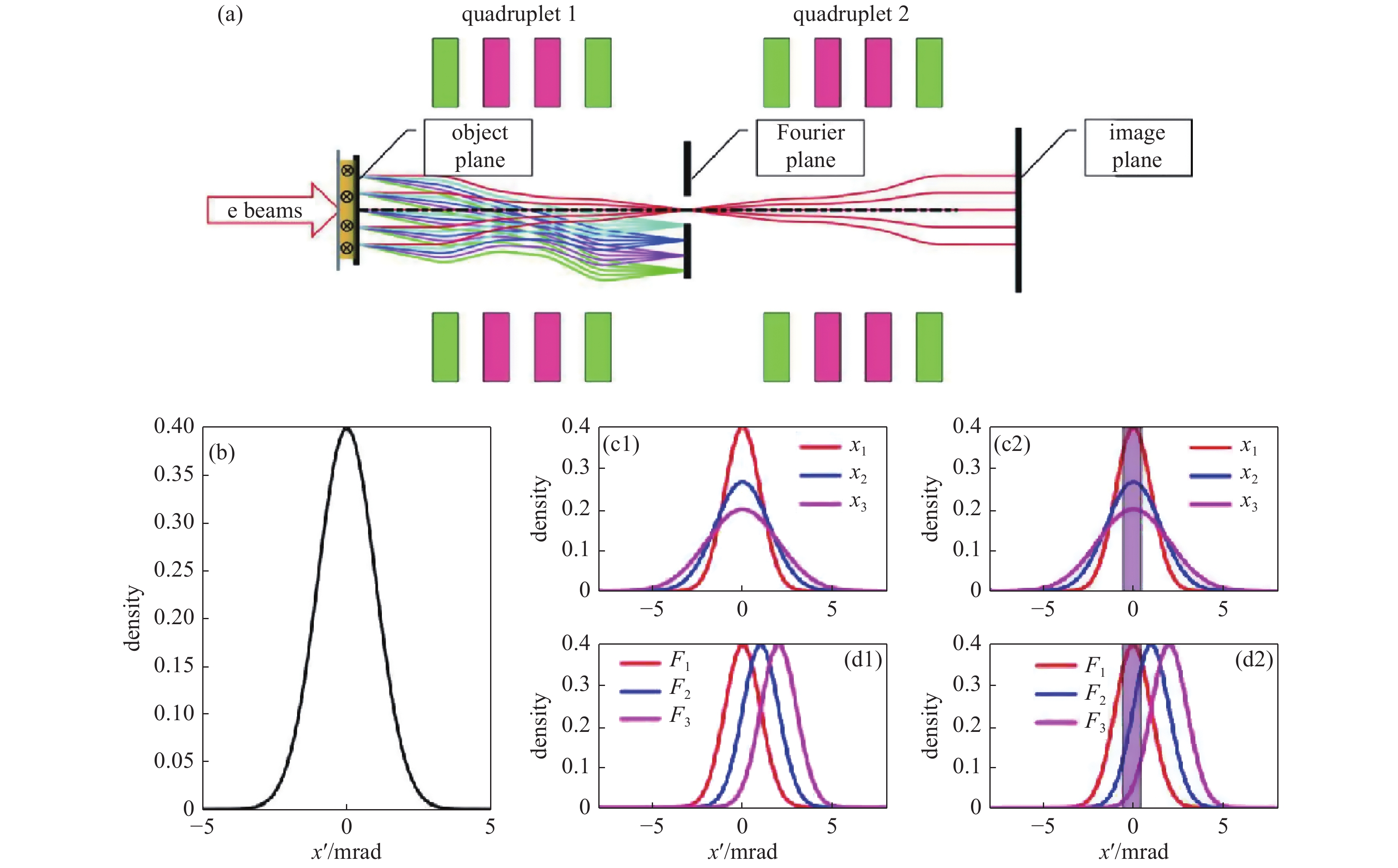
Figure 1. Schematic diagram of HEELR to make angle selection with an aperture. (a) The schematic of point-to-point imaging beamline with an aperture at the Fourier plane. (b) The angle distribution of incident electrons. (c1) and (c2) illustrate the electron angle distributions after target with different thicknesses and the corresponding position distributions at the Fourier plane. (d1) and (d2) are the electron angle distributions after penetrating the system with E/B field and the corresponding position distributions at the Fourier plane. The purple rectangular shadow indicates the aperture acceptance area
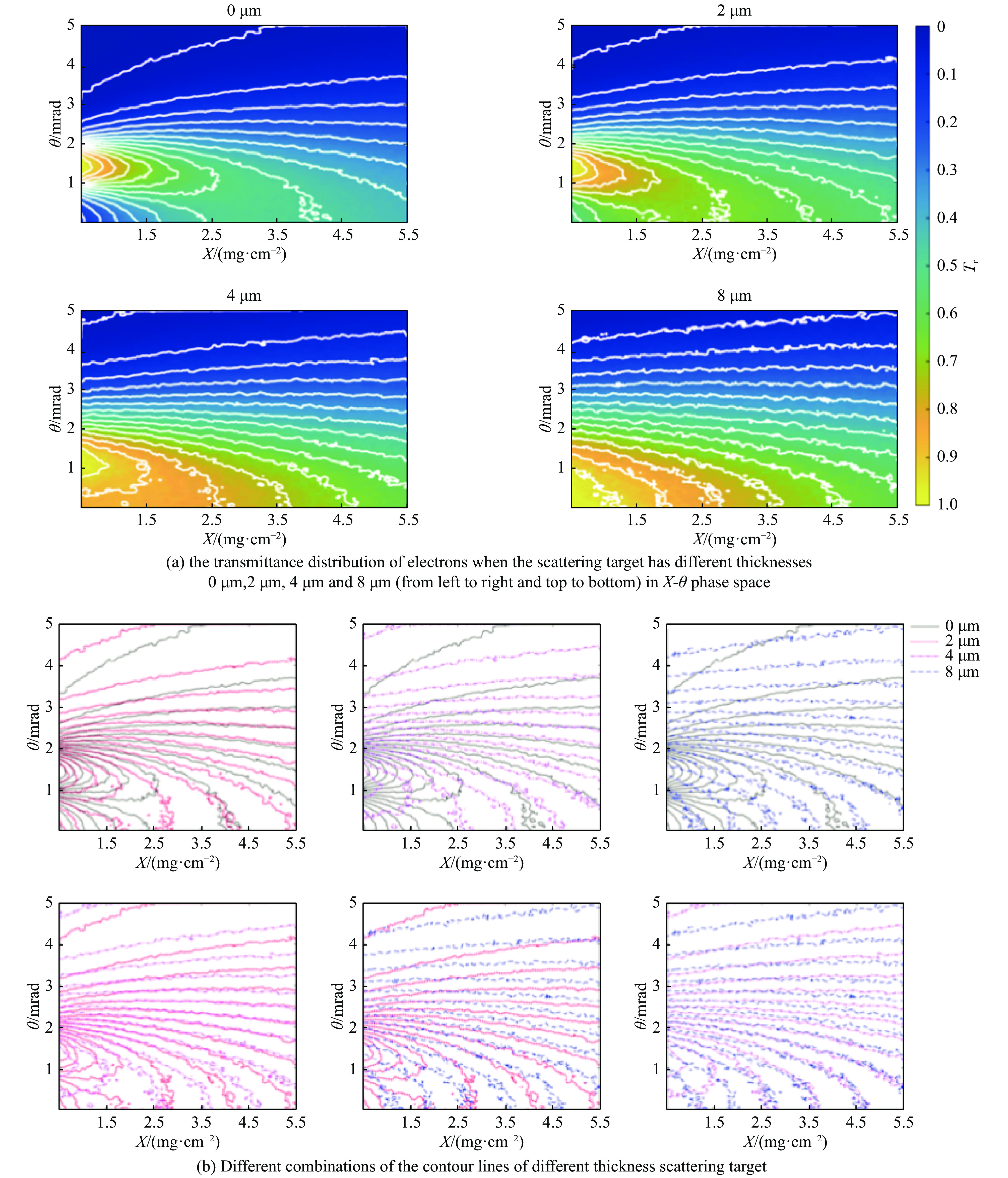
Figure 2. The principle of areal density difference method to diagnose the areal density and E/B field. (a) The scattering angle distributions after the grille scattering target. Different color corresponds to different thicknesses (t1 and t2). (b) The surface plot with contour lines of Tr (t+t1, θ) and Tr (t+t2, θ). The intersection point of two contour lines with the same transmittance marks the solution (t, θ)
Figure 3. Overall design of the dual degrees of freedom diagnostic. (a) The incident beams will be scattered by the scattering target before penetrating the real diagnosed target. (b) The front view (perpendicular to the beam bunches) of the scattering target, different color indicates different thickness
Figure 4. (a) The wedge shape represents the hydrogen sample with gradient areal density. The color indicates the value of deflection angle from the E/B field and the coordinate system is as shown. (b) The ellipse shape aperture sets the upper limit of the angle acceptance as 1mrad. (c) The ring shape aperture which sets the angle acceptance as (1,2) mrad
-
[1] Walsh C A, Chittenden J P, McGlinchey K, et al. Self-generated magnetic fields in the stagnation phase of indirect-drive implosions on the National Ignition Facility[J]. Physical Review Letters, 2017, 118: 155001. doi: 10.1103/PhysRevLett.118.155001 [2] Fox W, Bhattacharjee A, Germaschewski K. Magnetic reconnection in high-energy-density laser-produced plasmas[J]. Physics of Plasmas, 2012, 19: 056309. doi: 10.1063/1.3694119 [3] Gotchev O V, Chang P Y, Knauer J P, et al. Laser-driven magnetic-flux compression in high-energy-density plasmas[J]. Physical Review Letters, 2009, 103: 215004. doi: 10.1103/PhysRevLett.103.215004 [4] Lindemuth L R, Ekdahl C A, Fowler C M, et al. US/Russian collaboration in high-energy-density physics using high-explosive pulsed power: ultrahigh current experiments, ultrahigh magnetic field applications, and progress toward controlled thermonuclear fusion[J]. IEEE Transactions on Plasma Science, 1997, 25(6): 1357-1372. doi: 10.1109/27.650905 [5] Merrill F E, Campos E, Espinoza C, et al. Magnifying lens for 800 MeV proton radiography[J]. Review of Scientific Instruments, 2011, 82: 103709. doi: 10.1063/1.3652974 [6] Kantsyrev A V, Golubev A A, Bogdanov A V, et al. TWAC-ITEP proton microscopy facility[J]. Instruments and Experimental Techniques, 2014, 57(1): 1-10. doi: 10.1134/S0020441214010151 [7] Rygg J R, Séguin F H, Li C K, et al. Proton radiography of inertial fusion implosions[J]. Science, 2008, 319(5867): 1223-1225. doi: 10.1126/science.1152640 [8] Tommasini R, Landen O L, Hopkins L B, et al. Time-resolved fuel density profiles of the stagnation phase of indirect-drive inertial confinement implosions[J]. Physical Review Letters, 2020, 125: 155003. doi: 10.1103/PhysRevLett.125.155003 [9] Martynenko A S, Pikuz S A, Skobelev I Y, et al. Optimization of a laser plasma-based X-ray source according to WDM absorption spectroscopy requirements[J]. Matter and Radiation at Extremes, 2021, 6: 014405. doi: 10.1063/5.0025646 [10] Zhao Yongtao, Zhang Zimin, Gai Wei, et al. High energy electron radiography scheme with high spatial and temporal resolution in three dimension based on a e-LINAC[J]. Laser and Particle Beams, 2016, 34(2): 338-342. doi: 10.1017/S0263034616000124 [11] Xiao Jiahao, Zhang Zimin, Cao Shuchun, et al. Areal density and spatial resolution of high energy electron radiography[J]. Chinese Physics B, 2018, 27: 035202. doi: 10.1088/1674-1056/27/3/035202 [12] Wang Feng, Jiang Shaoen, Ding Yongkun, et al. Recent diagnostic developments at the 100 kJ-level laser facility in China[J]. Matter and Radiation at Extremes, 2020, 5: 035201. doi: 10.1063/1.5129726 [13] Li C K, Séguin F H, Frenje J A, et al. Measuring E and B fields in laser-produced plasmas with monoenergetic proton radiography[J]. Physical Review Letters, 2006, 97: 135003. doi: 10.1103/PhysRevLett.97.135003 [14] Li C K, Séguin F H, Frenje J A, et al. Monoenergetic-proton-radiography measurements of implosion dynamics in direct-drive inertial-confinement fusion[J]. Physical Review Letters, 2008, 100: 225001. doi: 10.1103/PhysRevLett.100.225001 [15] Liao Guoqian, Li Yutong, Zhu Baojun, et al. Proton radiography of magnetic fields generated with an open-ended coil driven by high power laser pulses[J]. Matter and Radiation at Extremes, 2016, 1(4): 187-191. doi: 10.1016/j.mre.2016.06.003 [16] Schumaker W, Nakanii N, McGuffey C, et al. Ultrafast electron radiography of magnetic fields in high-intensity laser-solid interactions[J]. Physical Review Letters, 2013, 110: 015003. doi: 10.1103/PhysRevLett.110.015003 [17] Zhu P F, Zhang Z C, Chen L, et al. Ultrashort electron pulses as a four-dimensional diagnosis of plasma dynamics[J]. Review of Scientific Instruments, 2010, 81: 103505. doi: 10.1063/1.3491994 [18] Li Junjie, Wang Xuan, Chen Zhaoyang, et al. Ultrafast electron beam imaging of femtosecond laser-induced plasma dynamics[J]. Journal of Applied Physics, 2010, 107: 083305. doi: 10.1063/1.3380846 [19] Chen Long, Li Runze, Chen Jie, et al. Mapping transient electric fields with picosecond electron bunches[J]. Proceedings of the National Academy of Sciences of the United States of America, 2015, 112(47): 14479-14483. doi: 10.1073/pnas.1518353112 [20] Merrill F, Harmon F, Hunt A, et al. Electron radiography[J]. Nuclear Instruments and Methods in Physics Research Section B: Beam Interactions with Materials and Atoms, 2007, 261(1/2): 382-386. [21] Merrill F E, Goett J, Gibbs J W, et al. Demonstration of transmission high energy electron microscopy[J]. Applied Physics Letters, 2018, 112: 144103. doi: 10.1063/1.5011198 [22] Zhou Zheng, Fang Yu, Chen Han, et al. Visualizing the melting processes in ultrashort intense laser triggered gold mesh with high energy electron radiography[J]. Matter and Radiation at Extremes, 2019, 4: 065402. doi: 10.1063/1.5109855 [23] Xiao Jiahao, Du Yingchao, Zhang Shizheng, et al. Ultrafast high-energy electron radiography application in magnetic field delicate structure measurement[J]. Laser and Particle Beams, 2021: 6683245. [24] Xiao Jiahao, Du Yingchao, Li Haoqing, et al. Ultrafast high energy electron lens radiography suitable for transient electromagnetic field diagnosis [J]. Journal of Instrumentation, 2022, 17(01):P01033 DOI: 10.1088/1748-0221/17/01/P01033 [25] Hirayama H, Namito Y, Bielajew A F, et al. The EGS5 code system[R]. SLAC-R-730, 2005. -


 下载:
下载:


