

Brief introduction of low-energy diffraction limited storage-ring-based synchrotron radiation and its applications
-
摘要: 在科学技术新需求的推动下,同步辐射光源持续往前发展。目前,同步辐射装置发展已历经三代,正处于第四代同步辐射光源蓬勃发展阶段。基于衍射极限储存环的同步辐射装置是第四代同步辐射光源的典型代表之一。第四代同步辐射光源主要发展趋势是进一步减小电子束流发射度,使光源具有极好的横向相干性,以及产生圆截面辐射的能力。如果束流发射度降至光学衍射极限“辐射波长/4π”,其亮度比第三代同步辐射光源高2个数量级。这种同步辐射光源在性能上发生的质的飞跃,将给同步辐射实验技术带来实质性的突破,从而给前沿科学技术研究和现代产业发展带来全新的机遇。从国际同步辐射发展趋势入手,首先介绍低能区衍射限储存环光源的特色和性能,然后介绍其带来的同步辐射实验技术的进步,并浅析低能区衍射限储存环光源在材料科学、能源科学、生命科学和环境科学上的应用,以及其带来的产业机遇。最后,总结和展望了低能区衍射限储存环光源带来的技术突破和潜在的应用前景。
-
关键词:
- 第四代同步辐射 /
- 低能区衍射极限储存环 /
- X射线实验技术 /
- 科学研究 /
- 产业机遇
Abstract: Driven by the demand of science and technology, synchrotron radiation (SR) facilities continue to develop. At present, the development of SR has gone through three generations, and is in the vigorous development stage of the fourth generation (4th). SR based on diffraction-limited storage ring is one of the typical representative of the 4th synchrotron light sources. The mainstream of the 4th SR is to further reduce the electron beam emittance, so that the light source from the 4th SR exhibits excellent transversal coherence and is able to produce circular cross-section radiation. If the beam emittance drops to the optical diffraction limited “radiation wavelength/4π”, its brightness enhances to 2-3 orders of magnitude higher than that of the third generation SR light source. The qualitative leap in the performance of this SR light source will bring a substantive breakthrough to the SR-based experimental techniques, and bring new opportunities to the cutting-edge scientific and technological research, as well as the development of modern industry. Starting with the development trend of worldwide SR facilities, this review first introduces the characteristics and performance of low-energy diffraction limited storage ring (DLSR) light source, then introduces the progress of experimental techniques brought by DLSR, and illustrates the application of low-energy DLSR light source in material science, energy science, life science and environmental science, as well as its industrial opportunities. Finally, the technical breakthrough and potential application prospect of low energy DLSR light source are summarized and prospected. -
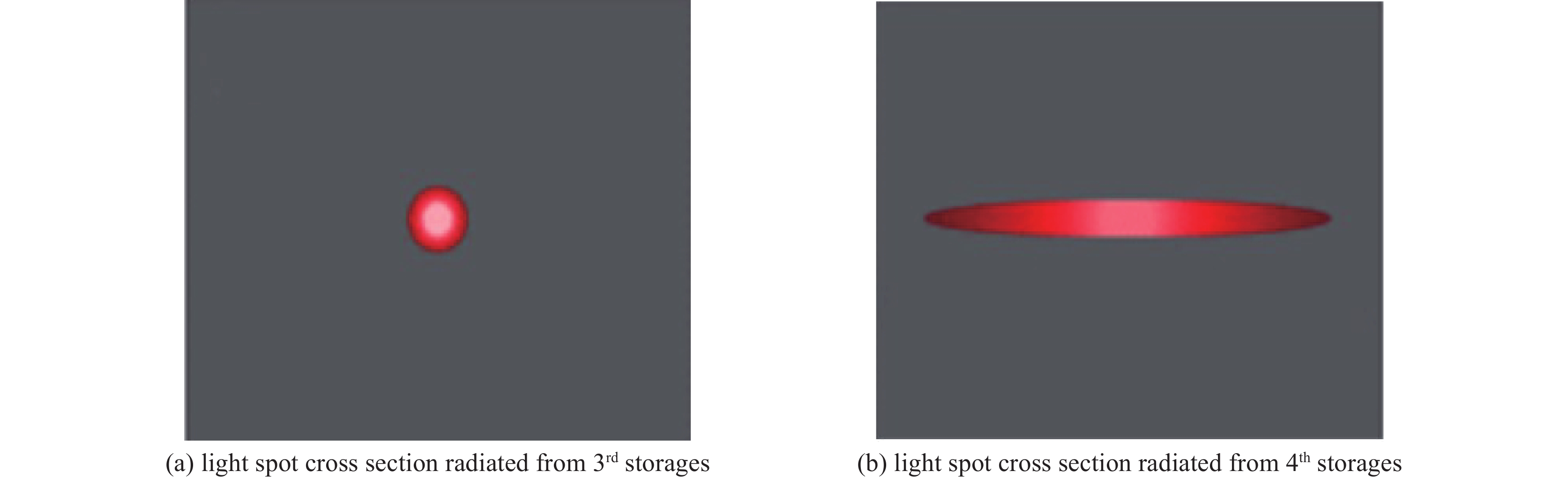
图 1 三代储存环光斑横截面与四代衍射限储存环光斑横截面的比较
Figure 1. Comparison of light spot cross section between 3rd generation storage rings and 4th generation diffraction-limited storage rings

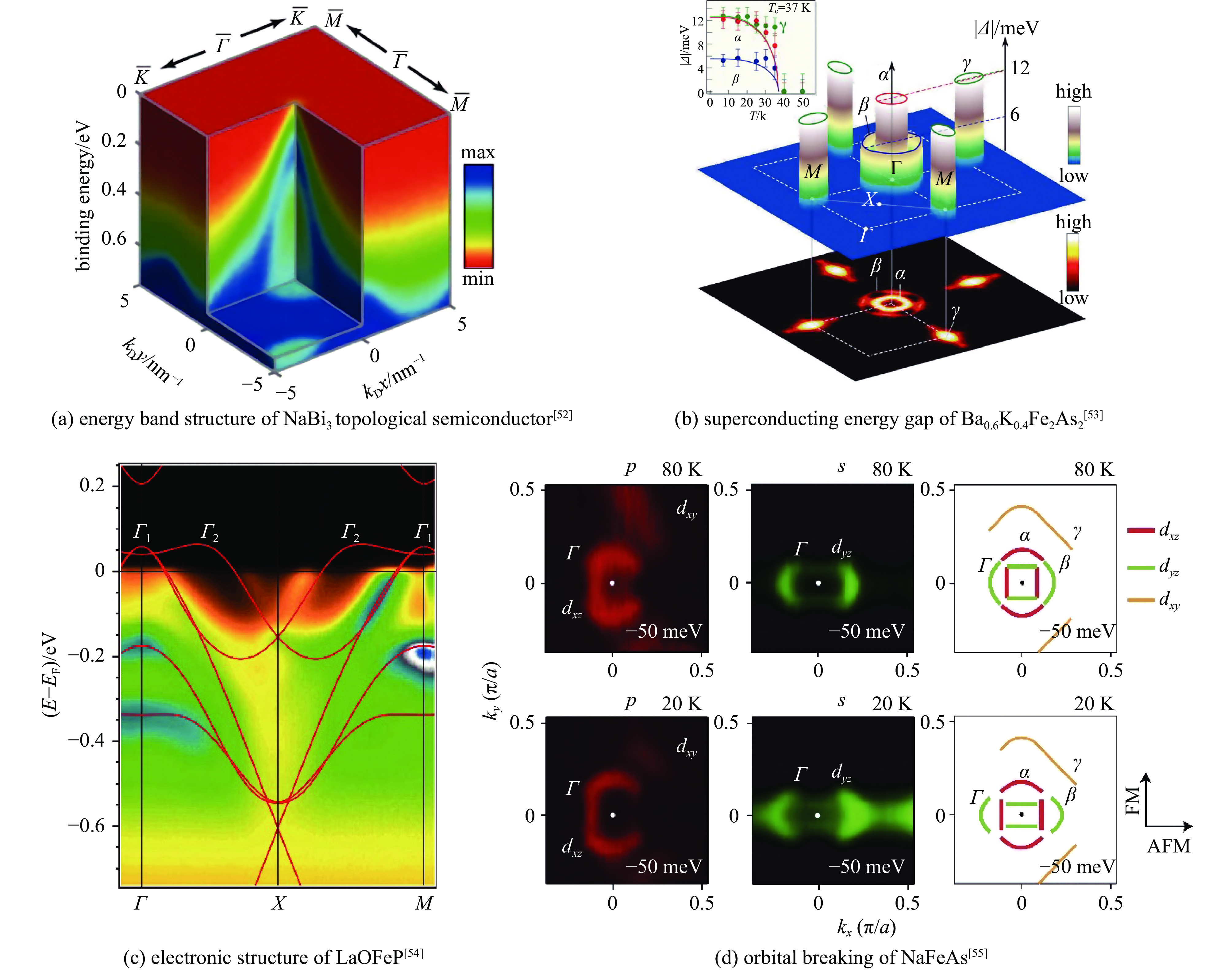
图 6 ARPES对量子材料的电子结构表征能力
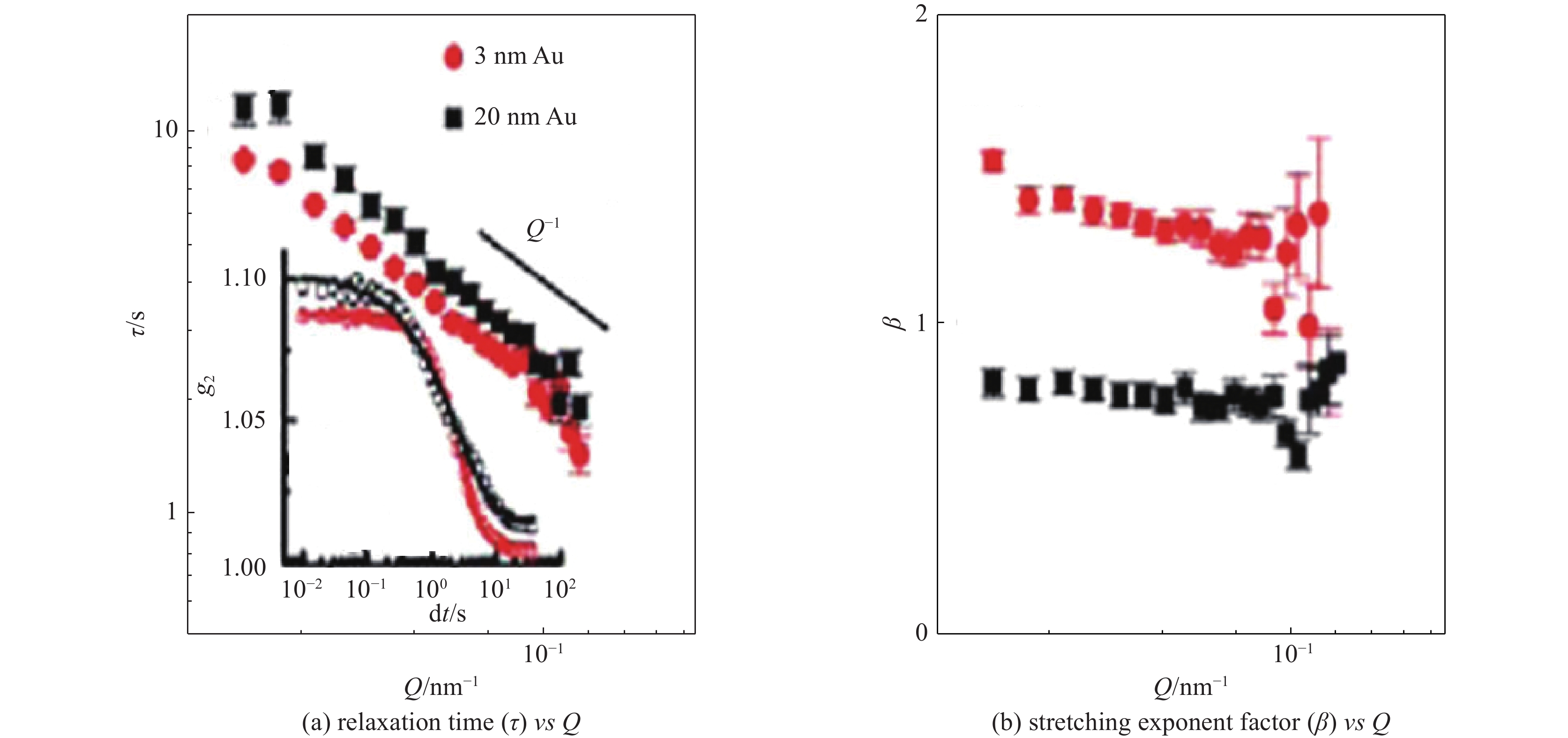
Figure 6. Characterization of electronic structure of quantum materials by ARPES
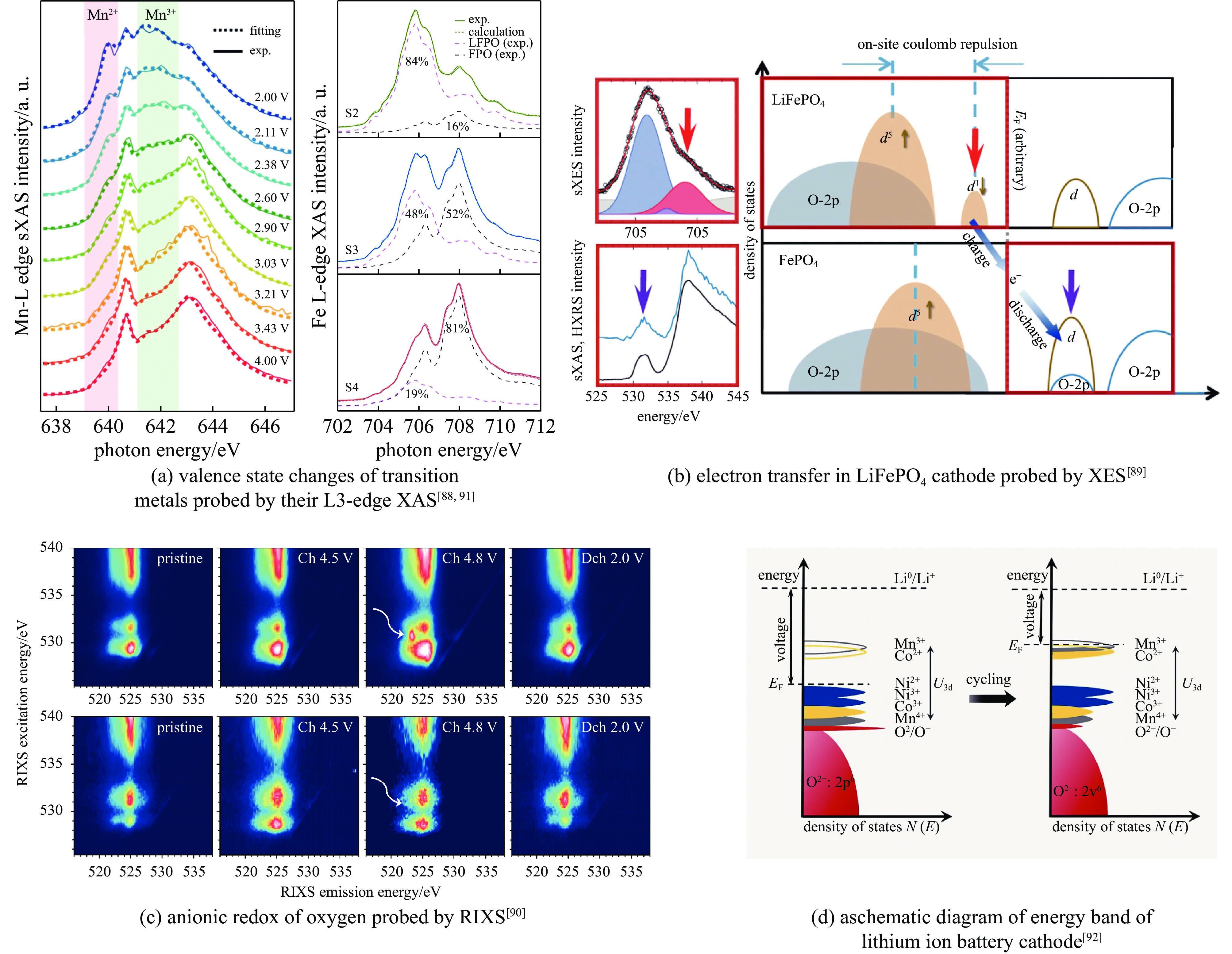
图 11 同步辐射装置提供全能带的X射线谱学表征技术,如sXAS、XES和RIXS等,描绘电极材料在充放电过程中的电子结构变化,指导材料合成和优化
Figure 11. SR lights provide versatile X-ray spectroscopic techniques, such as sXAS, XES and RIXS, to describe the electronic structure changes of electrode materials during charge and discharge, and guide the synthesis of materials and the optimization of their performance
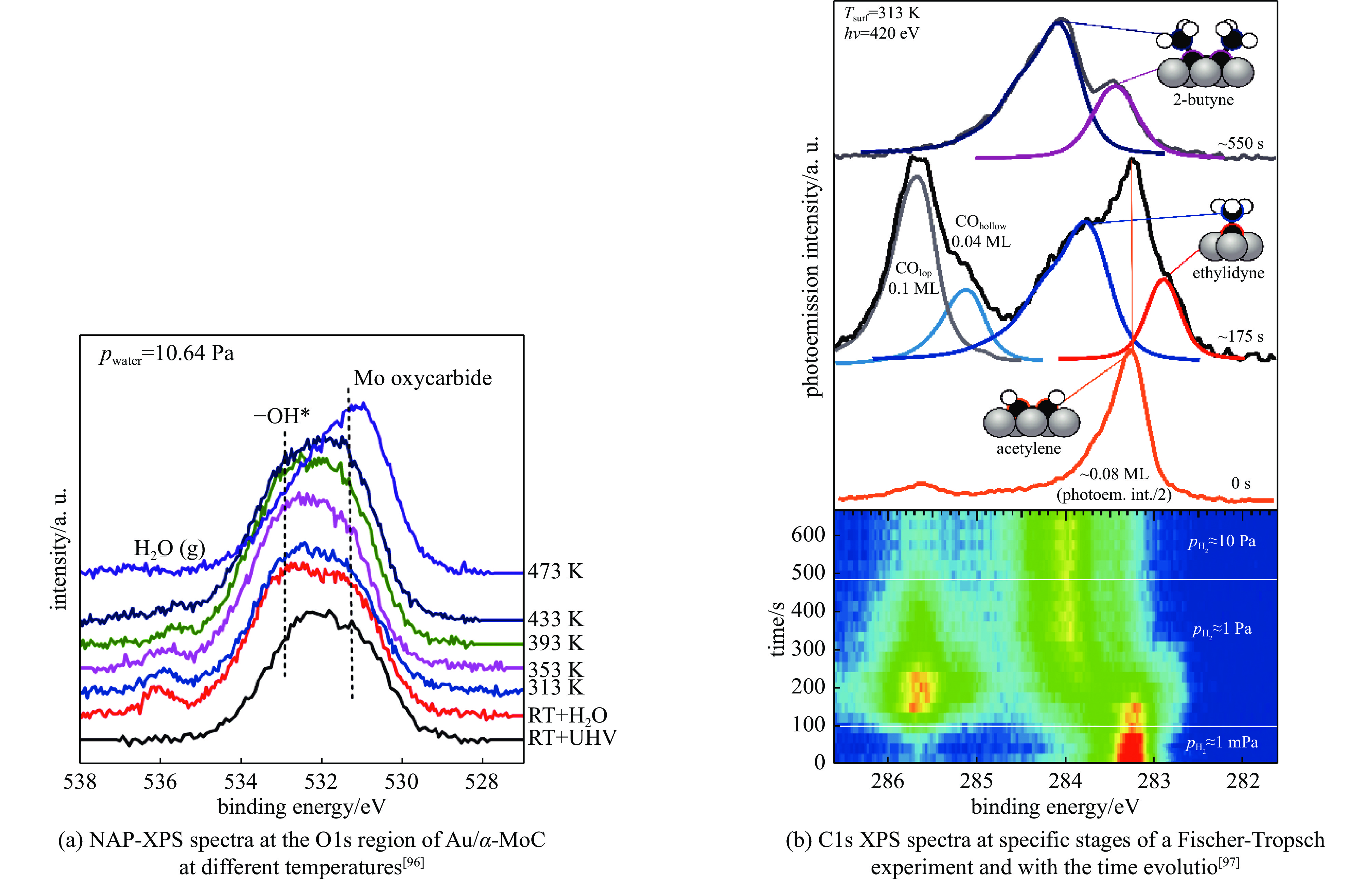
图 12 近常压XPS研究水煤气反应和费托合成反应机制
Figure 12. Near-ambient pressure XPS characterization for water-gas reaction and Fischer-Tropsch reaction
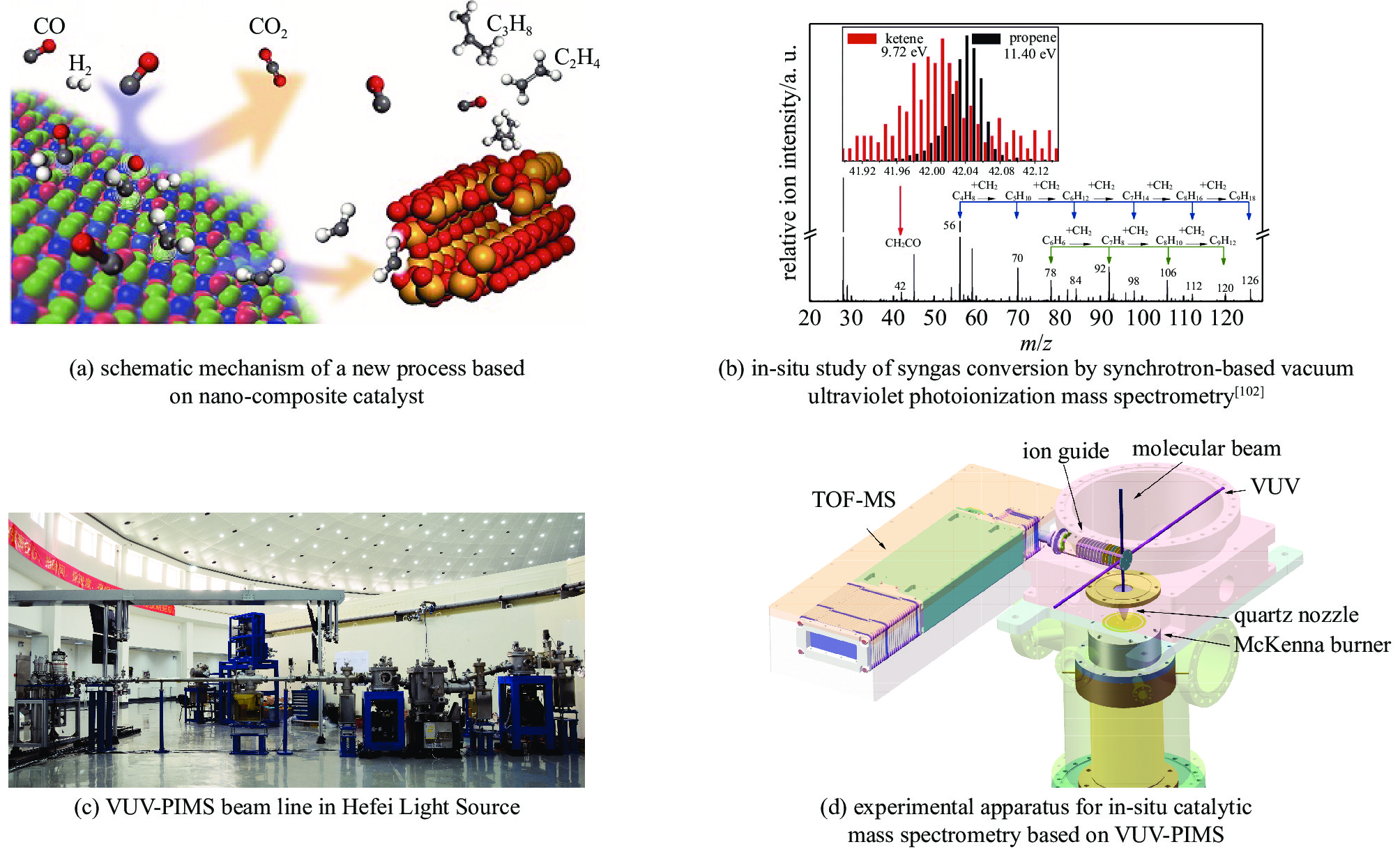
图 13 合肥光源原位催化质谱技术代表性研究结果
Figure 13. In-situ catalytic mass spectrometry in Hefei Light Source and its representative research results
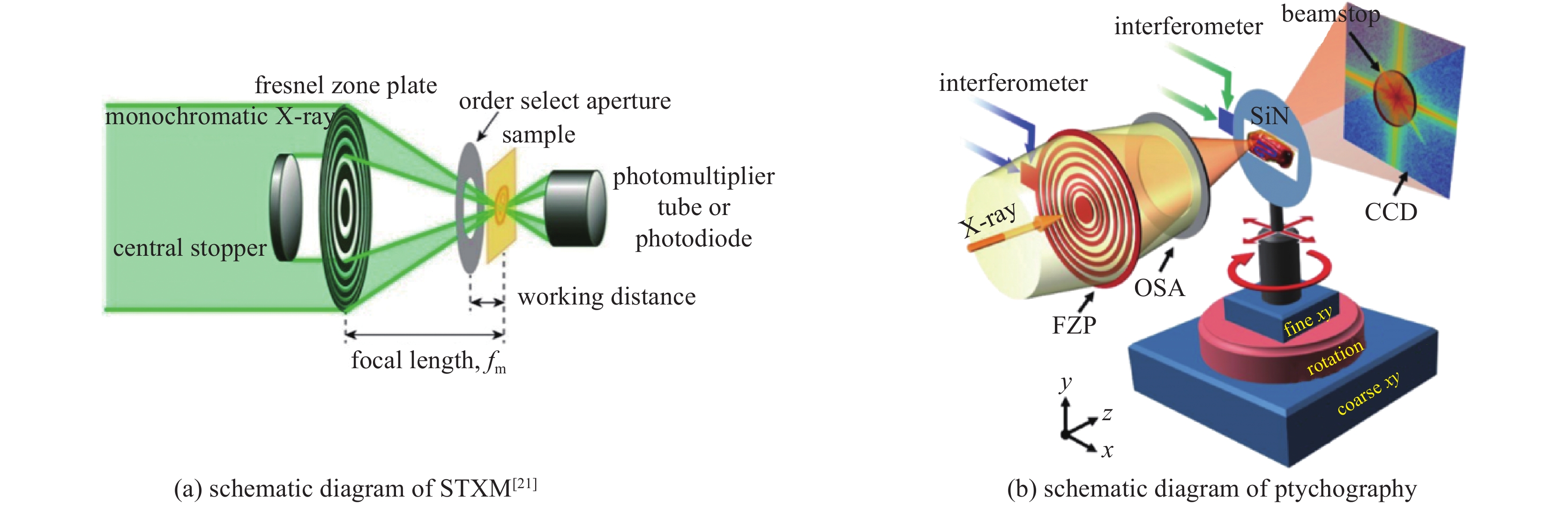
-
[1] Brown G S, Moncton D E. Handbook on synchrotron radiation[M]. New York: North-Holland, 1991. [2] 冼鼎昌. 神奇的光-同步辐射[M]. 长沙: 湖南教育出版社, 1994Xian Dingchang. Synchrtron radiation—the magic light[M]. Changsha: Hunan Education Publishing House, 1994 [3] Elder F R, Gurewitsch A M, Langmuir R V, et al. Radiation from electrons in a synchrotron[J]. Physical Review, 1947, 71(11): 829-830. [4] 姜晓明, 修立松. 同步辐射及其应用[M]. 北京: 北京科学技术出版社, 1996Jiang Xiaoming, Xiu Lisong. Synchrotron radiation and its applications[M]. Beijing: Beijing Science and Technology Press, 1996 [5] 马礼敦, 杨富家. 同步辐射应用概论[M]. 2版. 上海: 复旦大学出版社, 2005Ma Lidun, Yang Fujia. Introduction to synchrotron radiation applications[M]. 2nd ed. Shanghai: Fudan University Press, 2005. [6] 麦振洪. 同步辐射光的发展历史与现状——介绍新书《同步辐射光源及其应用》[J]. 现代物理知识, 2014, 26(2):65-71 doi: 10.13405/j.cnki.xdwz.2014.02.023Mai Zhenhong. Development history and current situation of synchrotron radiation—introduction to the new book “synchrotron radiation source and its application”[J]. Modern Physics, 2014, 26(2): 65-71 doi: 10.13405/j.cnki.xdwz.2014.02.023 [7] Hettel R. DLSR design and plans: an international overview[J]. Journal of Synchrotron Radiation, 2014, 21(5): 843-855. doi: 10.1107/S1600577514011515 [8] Hitchcock A P, Toney M F. Spectromicroscopy and coherent diffraction imaging: focus on energy materials applications[J]. Journal of Synchrotron Radiation, 2014, 21(5): 1019-1030. doi: 10.1107/S1600577514013046 [9] Frenkel A I, van Bokhoven J A. X-ray spectroscopy for chemical and energy sciences: the case of heterogeneous catalysis[J]. Journal of Synchrotron Radiation, 2014, 21(5): 1084-1089. doi: 10.1107/S1600577514014854 [10] de Jonge M D, Ryan C G, Jacobsen C J. X-ray nanoprobes and diffraction-limited storage rings: opportunities and challenges of fluorescence tomography of biological specimens[J]. Journal of Synchrotron Radiation, 2014, 21(5): 1031-1047. doi: 10.1107/S160057751401621X [11] Liu Feng, Brady M A, Wang Cheng. Resonant soft X-ray scattering for polymer materials[J]. European Polymer Journal, 2016, 81: 555-568. doi: 10.1016/j.eurpolymj.2016.04.014 [12] Comin R, Damascelli A. Resonant X-ray scattering studies of charge order in cuprates[J]. Annual Review of Condensed Matter Physics, 2016, 7: 369-405. doi: 10.1146/annurev-conmatphys-031115-011401 [13] Shpyrko O G. X-ray photon correlation spectroscopy[J]. Journal of Synchrotron Radiation, 2014, 21(5): 1057-1064. doi: 10.1107/S1600577514018232 [14] Sandy A R, Zhang Qingteng, Lurio L B. Hard X-ray photon correlation spectroscopy methods for materials studies[J]. Annual Review of Materials Research, 2018, 48: 167-190. doi: 10.1146/annurev-matsci-070317-124334 [15] Tamarat P, Bodnarchuk M I, Trebbia J B, et al. The ground exciton state of formamidinium lead bromide perovskite nanocrystals is a singlet dark state[J]. Nature Materials, 2019, 18(7): 717-724. doi: 10.1038/s41563-019-0364-x [16] Ehrburger-Dolle F, Morfin I, Bley F, et al. XPCS investigation of the dynamics of filler particles in stretched filled elastomers[J]. Macromolecules, 2012, 45(21): 8691-8701. doi: 10.1021/ma3013674 [17] Kukreja R, Hua N, Ruby J, et al. Orbital domain dynamics in magnetite below the Verwey transition[J]. Physical Review Letters, 2018, 121: 177601. doi: 10.1103/PhysRevLett.121.177601 [18] Chen X M, Thampy V, Mazzoli C, et al. Remarkable stability of charge density wave order in La1.875Ba0.125CuO4[J]. Physical Review Letters, 2016, 117: 167001. doi: 10.1103/PhysRevLett.117.167001 [19] Kirz J, Rarback H. Soft X-ray microscopes[J]. Review of Scientific Instruments, 1985, 56(1): 1-13. doi: 10.1063/1.1138464 [20] Pfeiffer F. X-ray ptychography[J]. Nature Photonics, 2018, 12(1): 9-17. doi: 10.1038/s41566-017-0072-5 [21] Ohigashi T, Yuzawa H, Kosugi N. A low-pass filtering Fresnel zone plate for soft X-ray microscopic analysis down to the lithium K-edge region[J]. Review of Scientific Instruments, 2020, 91: 103110. doi: 10.1063/5.0020956 [22] Chao Weilun, Harteneck B D, Liddle J A, et al. Soft X-ray microscopy at a spatial resolution better than 15 nm[J]. Nature, 2005, 435(7046): 1210-1213. doi: 10.1038/nature03719 [23] Shapiro D A, Yu Y S, Tyliszczak T, et al. Chemical composition mapping with nanometre resolution by soft X-ray microscopy[J]. Nature Photonics, 2014, 8(10): 765-769. doi: 10.1038/nphoton.2014.207 [24] Shi Xiaowen, Burdet N, Chen Bo, et al. X-ray ptychography on low-dimensional hard-condensed matter materials[J]. Applied Physical Reviews, 2019, 6: 011306. doi: 10.1063/1.5045131 [25] Liu Xiaosong, Yang Wanli, Liu Zhi. Recent progress on synchrotron-based in-situ soft X-ray spectroscopy for energy materials[J]. Advanced Materials, 2014, 26(46): 7710-7729. doi: 10.1002/adma.201304676 [26] Berkeley Lab, U. S. Department of Energy Office of Science, ALS-U. ALS-U: solving scientific challenges with coherent soft X-rays, workshop report on early science enabled by the Advanced Light Source Upgrade[R]. 2017. [27] Avila J, Asensio M C. First NanoARPES user facility available at SOLEIL: an innovative and powerful tool for studying advanced materials[J]. Synchrotron Radiation News, 2014, 27(2): 24-30. doi: 10.1080/08940886.2014.889549 [28] Kastl C, Koch R J, Chen C T, et al. Effects of defects on band structure and excitons in WS2 revealed by nanoscale photoemission spectroscopy[J]. ACS Nano, 2019, 13(2): 1284-1291. [29] Jia Chunjing, Wohlfeld K, Wang Yao, et al. Using RIXS to uncover elementary charge and spin excitations[J]. Physical Review X, 2016, 6: 021020. [30] Qiao Ruimin, Li Qinghao, Zhuo Zengqing, et al. High-efficiency in situ resonant inelastic X-ray scattering (iRIXS) endstation at the Advanced Light Source[J]. Review of Scientific Instruments, 2017, 88: 033106. doi: 10.1063/1.4977592 [31] Chuang Yide, Feng Xuefei, Glans-Suzuki P A, et al. A design of resonant inelastic X-ray scattering (RIXS) spectrometer for spatial- and time-resolved spectroscopy[J]. Journal of Synchrotron Radiation, 2020, 27(3): 695-707. doi: 10.1107/S1600577520004440 [32] Kaiser A M, Schöppner C, Römer F M, et al. Nano and picosecond magnetization dynamics of weakly coupled CoFe/Cr/NiFe trilayers studied by a multitechnique approach[J]. Physical Review B, 2011, 84: 134406. doi: 10.1103/PhysRevB.84.134406 [33] van der Laan G, Figueroa A I. X-ray magnetic circular dichroism—A versatile tool to study magnetism[J]. Coordination Chemistry Reviews, 2014, 277/278: 95-129. doi: 10.1016/j.ccr.2014.03.018 [34] 吴义政. 同步辐射X射线磁二色性在自旋电子学研究中的应用[J]. 物理, 2010, 39(6):406-415Wu Yizheng. Applications of X-ray magnetic dichroism in spintronics[J]. Wuli, 2010, 39(6): 406-415 [35] Mengotti E, Heyderman L J, Rodríguez A F, et al. Real-space observation of emergent magnetic monopoles and associated Dirac strings in artificial kagome spin ice[J]. Nature Physics, 2011, 7(1): 68-74. doi: 10.1038/nphys1794 [36] Zhao Tong, Scholl A, Zavaliche F, et al. Electrical control of antiferromagnetic domains in multiferroic BiFeO3 films at room temperature[J]. Nature Materials, 2006, 5(10): 823-829. doi: 10.1038/nmat1731 [37] Suchorski Y, Kozlov S M, Bespalov I, et al. The role of metal/oxide interfaces for long-range metal particle activation during CO oxidation[J]. Nature Materials, 2018, 17(6): 519-522. doi: 10.1038/s41563-018-0080-y [38] Shiino T, Oh S H, Haney P M, et al. Antiferromagnetic domain wall motion driven by spin-orbit torques[J]. Physical Review Letters, 2016, 117: 087203. doi: 10.1103/PhysRevLett.117.087203 [39] Litzius K, Lemesh I, Krüger B, et al. Skyrmion Hall effect revealed by direct time-resolved X-ray microscopy[J]. Nature Physics, 2017, 13(2): 170-175. doi: 10.1038/nphys4000 [40] Wang Wenbo, Ou Yunbo, Liu Chang, et al. Direct evidence of ferromagnetism in a quantum anomalous Hall system[J]. Nature Physics, 2018, 14(8): 791-795. doi: 10.1038/s41567-018-0149-1 [41] Jiang Wanjun, Upadhyaya P, Zhang Wei, et al. Blowing magnetic skyrmion bubbles[J]. Science, 2015, 349(6245): 283-286. doi: 10.1126/science.aaa1442 [42] Fukami S, Zhang Chaoliang, DuttaGupta S, et al. Magnetization switching by spin-orbit torque in an antiferromagnet-ferromagnet bilayer system[J]. Nature Materials, 2016, 15(5): 535-541. doi: 10.1038/nmat4566 [43] Zhou Jiadong, Lin Junhao, Huang Xiangwei, et al. A library of atomically thin metal chalcogenides[J]. Nature, 2018, 556(7701): 355-359. doi: 10.1038/s41586-018-0008-3 [44] Baltz V, Manchon A, Tsoi M, et al. Antiferromagnetic spintronics[J]. Reviews of Modern Physics, 2018, 90: 015005. doi: 10.1103/RevModPhys.90.015005 [45] Lesne E, Fu Yu, Oyarzun S, et al. Highly efficient and tunable spin-to-charge conversion through Rashba coupling at oxide interfaces[J]. Nature Materials, 2016, 15(12): 1261-1266. doi: 10.1038/nmat4726 [46] Dagotto E. Complexity in strongly correlated electronic systems[J]. Science, 2005, 309(5732): 257-262. doi: 10.1126/science.1107559 [47] Keimer B, Moore J E. The physics of quantum materials[J]. Nature Physics, 2017, 13(11): 1045-1055. doi: 10.1038/nphys4302 [48] Tokura Y, Kawasaki M, Nagaosa N. Emergent functions of quantum materials[J]. Nature Physics, 2017, 13(11): 1056-1068. doi: 10.1038/nphys4274 [49] Basov D N, Averitt R D, Hsieh D. Towards properties on demand in quantum materials[J]. Nature Materials, 2017, 16(11): 1077-1088. doi: 10.1038/nmat5017 [50] Borisenko S V, Evtushinsky D V, Liu Zhonghao, et al. Direct observation of spin–orbit coupling in iron-based superconductors[J]. Nature Physics, 2016, 12(4): 311-317. doi: 10.1038/nphys3594 [51] Lu Donghui, Vishik IM, Yi Ming, et al. Angle-resolved photoemission studies of quantum materials[J]. Annual Review of Condensed Matter Physics, 2012, 3: 129-167. doi: 10.1146/annurev-conmatphys-020911-125027 [52] Liu Z K, Zhou B, Zhang Yi, et al. Discovery of a three-dimensional topological dirac semimetal, Na3Bi[J]. Science, 2014, 343(6173): 864-867. doi: 10.1126/science.1245085 [53] Ding Hanjie, Richard P, Nakayama K, et al. Observation of Fermi-surface–dependent nodeless superconducting gaps in Ba0.6K0.4Fe2As2[J]. Europhysics Letters, 2008, 83: 47001. doi: 10.1209/0295-5075/83/47001 [54] Lu D H, Yi Ming, Mo S K, et al. Electronic structure of the iron-based superconductor LaOFeP[J]. Nature, 2008, 455(7209): 81-84. doi: 10.1038/nature07263 [55] Zhang Yong, He Cheng, Ye Z R, et al. Symmetry breaking via orbital-dependent reconstruction of electronic structure in detwinned NaFeAs[J]. Physical Review B, 2012, 85: 085121. doi: 10.1103/PhysRevB.85.085121 [56] Zhou Shuyun, Gweon GH, Fedorov AV, et al. Erratum: substrate-induced bandgap opening in epitaxial graphene[J]. Nature Materials, 2007, 6: 916. doi: 10.1038/nmat2056 [57] Fäth M, Freisem S, Menovsky A A, et al. Spatially inhomogeneous metal-insulator transition in doped manganites[J]. Science, 1999, 285(5433): 1540-1542. doi: 10.1126/science.285.5433.1540 [58] Song Canli, Wang Yilin, Cheng Peng, et al. Direct observation of nodes and twofold symmetry in FeSe superconductor[J]. Science, 2011, 332(6036): 1410-1413. doi: 10.1126/science.1202226 [59] Li Wei, Ding Hao, Deng Peng, et al. Phase separation and magnetic order in K-doped iron selenide superconductor[J]. Nature Physics, 2012, 8(2): 126-130. doi: 10.1038/nphys2155 [60] Parkin S. Racetrack memory: a storage class memory based on current controlled magnetic domain wall motion[C]//2009 Device Research Conference. 2009: 3-6. [61] Knafo W, Raymond S, Lejay P, et al. Antiferromagnetic criticality at a heavy-fermion quantum phase transition[J]. Nature Physics, 2009, 5(10): 753-757. doi: 10.1038/nphys1374 [62] Schröder A, Aeppli G, Coldea R, et al. Onset of antiferromagnetism in heavy-fermion metals[J]. Nature, 2000, 407(6802): 351-355. doi: 10.1038/35030039 [63] Duan Chunruo, Baumbach R E, Podlesnyak A, et al. Resonance from antiferromagnetic spin fluctuations for superconductivity in UTe2[J]. Nature, 2021, 600(7890): 636-640. doi: 10.1038/s41586-021-04151-5 [64] Shpyrko O G, Isaacs E D, Logan J M, et al. Direct measurement of antiferromagnetic domain fluctuations[J]. Nature, 2007, 447(7140): 68-71. doi: 10.1038/nature05776 [65] Kim K J, Kim S K, Hirata Y, et al. Fast domain wall motion in the vicinity of the angular momentum compensation temperature of ferrimagnets[J]. Nature Materials, 2017, 16(12): 1187-1192. doi: 10.1038/nmat4990 [66] Bernstein D P, Bräuer B, Kukreja R, et al. Nonuniform switching of the perpendicular magnetization in a spin-torque-driven magnetic nanopillar[J]. Physical Review B, 2011, 83: 180410. doi: 10.1103/PhysRevB.83.180410 [67] Reyren N, Bouzehouane K, Chauleau J Y, et al. Skyrmions in magnetic multilayers: chirality, electrical detection and current-induced motion[C]. Proceedings of SPIE, Spintronics X. 2017: 1035724. [68] Hellman F, Hoffmann A, Tserkovnyak Y, et al. Interface-induced phenomena in magnetism[J]. Reviews of Modern Physics, 2017, 89: 025006. doi: 10.1103/RevModPhys.89.025006 [69] Banerjee S, Erten O, Randeria M. Ferromagnetic exchange, spin-orbit coupling and spiral magnetism at the LaAlO3/SrTiO3 interface[J]. Nature Physics, 2013, 9(10): 626-630. doi: 10.1038/nphys2702 [70] Grisolia M N, Varignon J, Sanchez-Santolino G, et al. Hybridization-controlled charge transfer and induced magnetism at correlated oxide interfaces[J]. Nature Physics, 2016, 12(5): 484-492. doi: 10.1038/nphys3627 [71] Saito Y, Nakamura Y, Bahramy M S, et al. Superconductivity protected by spin-valley locking in ion-gated MoS2[J]. Nature Physics, 2016, 12(2): 144-149. doi: 10.1038/nphys3580 [72] Li J, Shelford L R, Shafer P, et al. Direct detection of pure ac spin current by X-ray pump-probe measurements[J]. Physical Review Letters, 2016, 117: 076602. doi: 10.1103/PhysRevLett.117.076602 [73] Li Wenjing, Bykova I, Zhang Shilei, et al. Anatomy of skyrmionic textures in magnetic multilayers[J]. Advanced Materials, 2019, 31: 1807683. doi: 10.1002/adma.201807683 [74] Tumbleston J R, Collins B A, Yang Liqiang, et al. The influence of molecular orientation on organic bulk heterojunction solar cells[J]. Nature Photonics, 2014, 8(5): 385-391. doi: 10.1038/nphoton.2014.55 [75] Collins B A, Cochran J E, Yan Hongping, et al. Polarized X-ray scattering reveals non-crystalline orientational ordering in organic films[J]. Nature Materials, 2012, 11(6): 536-543. doi: 10.1038/nmat3310 [76] Suh H S, Kang Huiman, Nealey P F, et al. Thickness dependence of neutral parameter windows for perpendicularly oriented block copolymer thin films[J]. Macromolecules, 2010, 43(10): 4744-4751. doi: 10.1021/ma100150j [77] Ahn H, Shin C, Lee B, et al. Phase transitions of block copolymer film on homopolymer-grafted substrate[J]. Macromolecules, 2010, 43(4): 1958-1963. doi: 10.1021/ma9022229 [78] Sivaniah E, Hayashi Y, Matsubara S, et al. Symmetric diblock copolymer thin films on rough substrates. Kinetics and structure formation in pure block copolymer thin films[J]. Macromolecules, 2005, 38(5): 1837-1849. doi: 10.1021/ma0482157 [79] Hur S M, Khaira G S, Ramírez-Hernández A, et al. Simulation of defect reduction in block copolymer thin films by solvent annealing[J]. ACS Macro Letters, 2015, 4(1): 11-15. doi: 10.1021/mz500705q [80] Sinturel C, Vayer M, Morris M, et al. Solvent vapor annealing of block polymer thin films[J]. Macromolecules, 2013, 46(14): 5399-5415. doi: 10.1021/ma400735a [81] De Rosa C, Park C, Thomas E L, et al. Microdomain patterns from directional eutectic solidification and epitaxy[J]. Nature, 2000, 405(6785): 433-437. doi: 10.1038/35013018 [82] Tang Chuanbing, Wu Wei, Smilgies D M, et al. Robust control of microdomain orientation in thin films of block copolymers by zone casting[J]. Journal of the American Chemical Society, 2011, 133(30): 11802-11809. doi: 10.1021/ja204724h [83] Saito I, Miyazaki T, Yamamoto K. Depth-resolved structure analysis of cylindrical microdomain in block copolymer thin film by grazing-incidence small-angle X-ray scattering utilizing low-energy x-rays[J]. Macromolecules, 2015, 48(22): 8190-8196. doi: 10.1021/acs.macromol.5b01883 [84] Gann E, Watson A, Tumbleston J R, et al. Topographic measurement of buried thin-film interfaces using a grazing resonant soft X-ray scattering technique[J]. Physical Review B, 2014, 90: 245421. doi: 10.1103/PhysRevB.90.245421 [85] Leheny R L. XPCS: nanoscale motion and rheology[J]. Current Opinion in Colloid & Interface Science, 2012, 17(1): 3-12. [86] Lu Jun, Wu Tianpin, Amine K. State-of-the-art characterization techniques for advanced lithium-ion batteries[J]. Nature Energy, 2017, 2: 17011. doi: 10.1038/nenergy.2017.11 [87] Peled E, Menkin S. Review—SEI: past, present and future[J]. Journal of the Electrochemical Society, 2017, 164(7): A1703-A1719. doi: 10.1149/2.1441707jes [88] Liu Xiaosong, Liu Jun, Qiao Ruimin, et al. Phase transformation and lithiation effect on electronic structure of LixFePO4: an in-depth study by soft X-ray and simulations[J]. Journal of the American Chemical Society, 2012, 134(33): 13708-13715. doi: 10.1021/ja303225e [89] Liu Xiaosong, Wang Y J, Barbiellini B, et al. Why LiFePO4 is a safe battery electrode: Coulomb repulsion induced electron-state reshuffling upon lithiation[J]. Physical Chemistry Chemical Physics, 2015, 17(39): 26369-26377. doi: 10.1039/C5CP04739K [90] Xu Jing, Sun Meiling, Qiao Ruimin, et al. Elucidating anionic oxygen activity in lithium-rich layered oxides[J]. Nature Communications, 2018, 9: 947. doi: 10.1038/s41467-018-03403-9 [91] Wu Jinpeng, Shen Zhixun, Yang Wanli. Redox mechanism in Na-ion battery cathodes probed by advanced soft X-ray spectroscopy[J]. Frontiers in Chemistry, 2020, 8: 816. doi: 10.3389/fchem.2020.00816 [92] Zhao Shuoqing, Yan Kang, Zhang Jinqiang, et al. Reaction mechanisms of layered lithium-rich cathode materials for high-energy lithium-ion batteries[J]. Angewandte Chemie International Edition, 2021, 60(5): 2208-2220. doi: 10.1002/anie.202000262 [93] House R A, Maitra U, Pérez-Osorio M A, et al. Superstructure control of first-cycle voltage hysteresis in oxygen-redox cathodes[J]. Nature, 2020, 577(7791): 502-508. doi: 10.1038/s41586-019-1854-3 [94] Yang Chunpeng, Fu Kun, Zhang Ying, et al. Protected lithium-metal anodes in batteries: from liquid to solid[J]. Advanced Materials, 2017, 29: 1701169. doi: 10.1002/adma.201701169 [95] Li Yiyang, Weker J N, Gent W E, et al. Dichotomy in the lithiation pathway of ellipsoidal and platelet LiFePO4 particles revealed through nanoscale operando state-of-charge imaging[J]. Advanced Functional Materials, 2015, 25(24): 3677-3687. doi: 10.1002/adfm.201500286 [96] Yao Siyu, Zhang Xiao, Zhou Wu, et al. Atomic-layered Au clusters on α-MoC as catalysts for the low-temperature water-gas shift reaction[J]. Science, 2017, 357(6349): 389-393. doi: 10.1126/science.aah4321 [97] Weststrate C J, Sharma D, Rodriguez D G, et al. Mechanistic insight into carbon-carbon bond formation on cobalt under simulated Fischer-Tropsch synthesis conditions[J]. Nature Communications, 2020, 11: 750. doi: 10.1038/s41467-020-14613-5 [98] Zou Xiaoxin, Zhang Yu. Noble metal-free hydrogen evolution catalysts for water splitting[J]. Chemical Society Reviews, 2015, 44(15): 5148-5180. doi: 10.1039/C4CS00448E [99] Favaro M, Yang Jinhui, Nappini S, et al. Understanding the oxygen evolution reaction mechanism on CoOx using Operando ambient-pressure X-ray photoelectron spectroscopy[J]. Journal of the American Chemical Society, 2017, 139(26): 8960-8970. doi: 10.1021/jacs.7b03211 [100] Su Xiaozhi, Wang Yu, Zhou Jing, et al. Operando spectroscopic identification of active sites in NiFe Prussian blue analogues as electrocatalysts: activation of oxygen atoms for oxygen evolution reaction[J]. Journal of the American Chemical Society, 2018, 140(36): 11286-11292. doi: 10.1021/jacs.8b05294 [101] Ma Qiuyu, Hu Chengyi, Liu Kunlong, et al. Identifying the electrocatalytic sites of nickel disulfide in alkaline hydrogen evolution reaction[J]. Nano Energy, 2017, 41: 148-153. doi: 10.1016/j.nanoen.2017.09.036 [102] Jiao Feng, Li Jinjing, Pan Xiulian, et al. Selective conversion of syngas to light olefins[J]. Science, 2016, 351(6277): 1065-1068. doi: 10.1126/science.aaf1835 [103] Cheng Kang, Zhou Wei, Kang Jincan, et al. Bifunctional catalysts for one-step conversion of syngas into aromatics with excellent selectivity and stability[J]. Chem, 2017, 3(2): 334-347. doi: 10.1016/j.chempr.2017.05.007 [104] Akri M, Zhao Shu, Li Xiaoyu, et al. Atomically dispersed nickel as coke-resistant active sites for methane dry reforming[J]. Nature Communications, 2019, 10: 5181. doi: 10.1038/s41467-019-12843-w [105] Li Xiaodong, Liang Liang, Sun Yongfu, et al. Ultrathin conductor enabling efficient IR light CO2 reduction[J]. Journal of the American Chemical Society, 2019, 141(1): 423-430. doi: 10.1021/jacs.8b10692 [106] Li Xiaodong, Sun Yongfu, Xu Jiaqi, et al. Selective visible-light-driven photocatalytic CO2 reduction to CH4 mediated by atomically thin CuIn5S8 layers[J]. Nature Energy, 2019, 4(8): 690-699. doi: 10.1038/s41560-019-0431-1 [107] Walker J E. ATP synthesis by rotary catalysis (Nobel lecture)[J]. Angewandte Chemie International Edition, 1998, 37(17): 2308-2319. doi: 10.1002/(SICI)1521-3773(19980918)37:17<2308::AID-ANIE2308>3.0.CO;2-W [108] MacKinnon R. Potassium channels and the atomic basis of selective ion conduction (Nobel lecture)[J]. Angewandte Chemie International Edition, 2004, 43(33): 4265-4277. doi: 10.1002/anie.200400662 [109] Kornberg R. The molecular basis of eukaryotic transcription (Nobel lecture)[J]. Angewandte Chemie International Edition, 2007, 46(37): 6956-6965. doi: 10.1002/anie.200701832 [110] Yonath A. Hibernating bears, antibiotics, and the evolving ribosome (Nobel lecture)[J]. Angewandte Chemie International Edition, 2010, 49(26): 4340-4354. doi: 10.1002/anie.201001297 [111] Kobilka B. The structural basis of G-protein-coupled receptor signaling (Nobel lecture)[J]. Angewandte Chemie International Edition, 2013, 52(25): 6380-6388. doi: 10.1002/anie.201302116 [112] Wang Liming, Zhang Tianlu, Li Panyun, et al. Use of synchrotron radiation-analytical techniques to reveal chemical origin of silver-nanoparticle cytotoxicity[J]. Acs Nano, 2015, 9(6): 6532-6547. doi: 10.1021/acsnano.5b02483 [113] Martins A C, Morcillo P, Ijomone O M, et al. New insights on the role of manganese in Alzheimer's disease and Parkinson's disease[J]. International Journal of Environmental Research and Public Health, 2019, 16: 3546. doi: 10.3390/ijerph16193546 [114] Dang Zheng, Guan Yong, Wu Zhao, et al. Regulating the synthesis rate and yield of bio-assembled FeS nanoparticles for efficient cancer therapy[J]. Nanoscale, 2021, 13(45): 18977-18986. doi: 10.1039/D1NR03591F [115] Coburn D S, Nazaretski E, Xu Weihe, et al. Design, characterization, and performance of a hard X-ray transmission microscope at the National Synchrotron Light Source II 18-ID beamline[J]. Review of Scientific Instruments, 2019, 90: 053701. doi: 10.1063/1.5088124 [116] Tang M T, Song Y F, Yin G C, et al. Hard X-ray microscopy with sub 30 nm spatial resolution[J]. AIP Conference Proceedings, 2007, 879(1): 1274-1277. [117] Chao Weilun, Fischer P, Tyliszczak T, et al. Real space soft X-ray imaging at 10 nm spatial resolution[J]. Optics Express, 2012, 20(9): 9777-9783. doi: 10.1364/OE.20.009777 [118] Miao Jianwei, Charalambous P, Kirz J, et al. Extending the methodology of X-ray crystallography to allow imaging of micrometre-sized non-crystalline specimens[J]. Nature, 1999, 400(6742): 342-344. doi: 10.1038/22498 [119] Huang Rujin, Zhang Yanlin, Bozzetti C, et al. High secondary aerosol contribution to particulate pollution during haze events in China[J]. Nature, 2014, 514(7521): 218-222. doi: 10.1038/nature13774 [120] Yao Lei, Garmash O, Bianchi F, et al. Atmospheric new particle formation from sulfuric acid and amines in a Chinese megacity[J]. Science, 2018, 361(6399): 278-281. doi: 10.1126/science.aao4839 [121] Johansson K O, Head-Gordon M P, Schrader P E, et al. Resonance-stabilized hydrocarbon-radical chain reactions may explain soot inception and growth[J]. Science, 2018, 361(6406): 997-1000. doi: 10.1126/science.aat3417 [122] Taatjes C A, Welz O, Eskola A J, et al. Direct measurements of conformer-dependent reactivity of the criegee intermediate CH3CHOO[J]. Science, 2013, 340(6129): 177-180. doi: 10.1126/science.1234689 [123] 刘义鹤, 江洪. 5G通信新材料研究进展[J]. 新材料产业, 2019(8):51-53 doi: 10.19599/j.issn.1008-892x.2019.08.013Liu Yihe, Jiang Hong. Research progress of new materials for 5G communication[J]. Advanced Materials Industry, 2019(8): 51-53 doi: 10.19599/j.issn.1008-892x.2019.08.013 [124] Manaila-Maximean D. Effective permittivity of a multi-phase system: nanoparticle-doped polymer-dispersed liquid crystal films[J]. Molecules, 2021, 26: 1441. doi: 10.3390/molecules26051441 [125] 师文钊, 刘瑾姝, 邢建伟, 等. 聚乙烯醇基相变复合材料研究进展[J]. 中国材料进展, 2020, 39(3):234-242 doi: 10.7502/j.issn.1674-3962-201902015Shi Wenzhao, Liu Jinshu, Xing Jianwei, et al. Research progress on polyvinyl alcohol-based phase change composites[J]. Materials China, 2020, 39(3): 234-242 doi: 10.7502/j.issn.1674-3962-201902015 [126] Hong G, Gan Xuemin, Leonhardt C, et al. A brief history of OLEDs-emitter development and industry milestones[J]. Advanced Materials, 2021, 33: 2005630. doi: 10.1002/adma.202005630 [127] Senses E, Ansar S M, Kitchens C L, et al. Small particle driven chain disentanglements in polymer nanocomposites[J]. Physical Review Letters, 2017, 118: 147801. doi: 10.1103/PhysRevLett.118.147801 [128] Wang Maoyu, Árnadóttir L, Xu Z J, et al. In situ X-ray absorption spectroscopy studies of nanoscale electrocatalysts[J]. Nano-Micro Letters, 2019, 11: 47. doi: 10.1007/s40820-019-0277-x [129] 刘涛, 纪军, 卫海桥, 等. 先进发动机燃烧基础研究的进展和关键科学问题[J]. 中国科学基金, 2014, 28(1):20-25 doi: 10.16262/j.cnki.1000-8217.2014.01.004Liu Tao, Ji Jun, Wei Haiqiao, et al. Progress and key scientific issues on advanced engine combustion research—summary of the 92nd Shuangqing forum of NFSC[J]. Bulletin of National Natural Science Foundation of China, 2014, 28(1): 20-25 doi: 10.16262/j.cnki.1000-8217.2014.01.004 [130] Dryer F L. Chemical kinetic and combustion characteristics of transportation fuels[J]. Proceedings of the Combustion Institute, 2015, 35(1): 117-144. doi: 10.1016/j.proci.2014.09.008 [131] Qi Fei. Combustion chemistry probed by synchrotron VUV photoionization mass spectrometry[J]. Proceedings of the Combustion Institute, 2013, 34(1): 33-63. doi: 10.1016/j.proci.2012.09.002 [132] Johansson K O, Dillstrom T, Monti M, et al. Formation and emission of large furans and oxygenated hydrocarbons from flames[J]. Proceedings of the National Academy of Sciences of the United States of America, 2016, 113(30): 8374-8379. doi: 10.1073/pnas.1604772113 [133] Battin-Leclerc F, Herbinet O, Glaude P A, et al. Experimental confirmation of the low-temperature oxidation scheme of alkanes[J]. Angewandte Chemie International Edition, 2010, 49(18): 3169-3172. doi: 10.1002/anie.200906850 [134] Yang Bin, Oßwald P, Li Yuyang, et al. Identification of combustion intermediates in isomeric fuel-rich premixed butanol–oxygen flames at low pressure[J]. Combustion and Flame, 2007, 148(4): 198-209. doi: 10.1016/j.combustflame.2006.12.001 [135] Taatjes C A, Hansen N, McIlroy A, et al. Enols are common intermediates in hydrocarbon oxidation[J]. Science, 2005, 308(5730): 1887-1889. doi: 10.1126/science.1112532 [136] Qi F, Li Y Y, et al. The second prize in National Natural Science Awards. 2018. -

 下载:
下载:
 点击查看大图
计量
- 文章访问数: 1915
- HTML全文浏览量: 586
- PDF下载量: 217
- 被引次数: 0
