

Source-coded radiography technique with high spatial-resolution for X-ray source driven by ps-laser
-
摘要: 为实现惯性约束聚变(ICF)内爆燃烧停滞阶段过程中最大压缩时刻的冷燃料面密度分布测量,设计了包含字母客体与针孔阵列的照相客体,通过同一发相同视角测量源分布与客体照相技术,首次建立了皮秒激光驱动的高能X射线源编码照相技术。通过星光III实验研究,基于W丝阵靶照相的反演图像空间分辨率5.4 μm±0.7 μm;激光到X射线(50~200 keV)的能量转换效率,W丝阵靶5.4×10⁻⁴,与传统Au单丝靶的转换效率(4.8×10⁻⁴)一致。基于源编码照相解决了传统皮秒激光背光照相中空间分辨率与光源亮度不能兼顾的困难,为强背景干扰下提供高信噪比、高分辨率的ICF靶丸压缩背光图像提供了重要照相方式。Abstract: To measure the areal density distribution of cold fuel at the maximum compression time during the stagnation phase of implosion in inertial confinement fusion (ICF), we have established the ps-laser driven high-energy X-ray radiography using source-coded technique. This paper describes the design and employment of the object including character-object and pinhole array. Based on the object, the source distribution and the object radiography was obtained at the same shot and same angle of view, and therefore the source-coded radiography of ps-laser driven X-ray has been established in experiments for the first time. From the experimental work on Xingguang-III facility, the spatial resolution of the inversion image with W wire-array target is 5.4 μm±0.7 μm. The efficiency of converting laser energy to high-energy bremsstrahlung (50−200 keV) is 5.4×10⁻⁴ in W wire-array target and 4.8×10⁻⁴ in Au single-wire target, respectively. It is possible that the the source-coded radiography of ps-laser driven X-ray in this work could account for overcoming the balance between spatial resolution and brightness in traditional X-ray backlight by ps-laser. The source-coded radiography provides an important method for ICF implosion backlight to get high resolution high signal-to-noise ratio images under the strong background.
-
Key words:
- inertial confinement fusion /
- ps laser /
- backlight /
- high-energy X-ray /
- spatial resolution
-

图 4 不同光源亮度下模拟半影图像与对应重建源图像
Figure 4. Simulated penumbral image and corresponding reconstructed source image at different X-ray brightness

图 5 不同辐射能谱下单丝照相图像
Figure 5. Radiography image by single-wire target with different X-ray spectrum

图 6 不同辐射能谱下丝阵靶模拟结果对应的反演图像
Figure 6. Inversion image from simulated image by wire-array target with different X-ray spectrum

图 8 客体设计图与实验照相图像
Figure 8. Design drawing of object and experimental radiography image

图 10 丝阵靶的针孔图像、半影图像与半影重建源图像
Figure 10. Pinhole image, penumbral image and reconstructed image from penumbral image of wire-array target
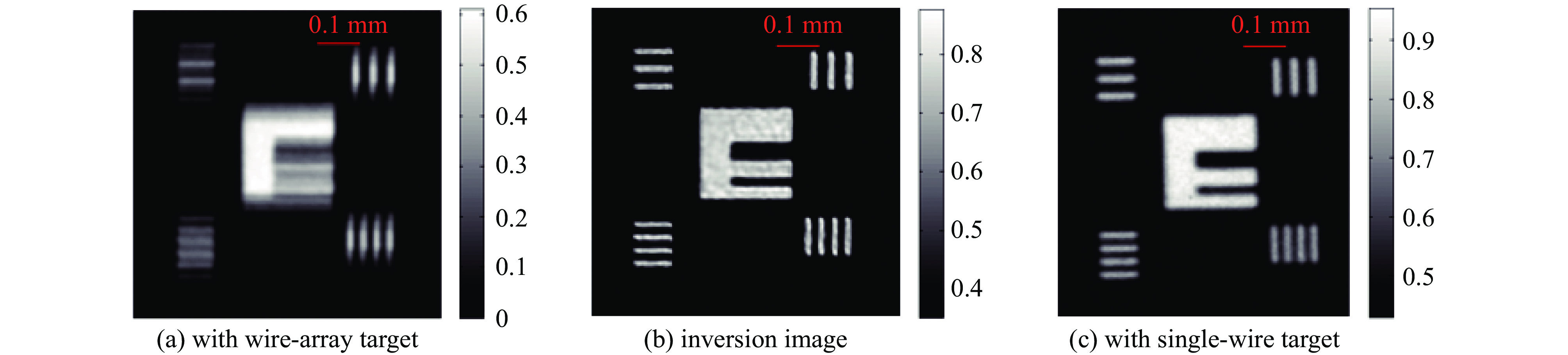
图 11 丝阵靶的照相图像、反演图像和单丝靶照相图像
Figure 11. Radiography image with wire-array target, inversion image and radiography image with single-wire target

图 12 丝阵靶反演图像和单丝靶照相图像的边缘扩散曲线
Figure 12. Edge spread curves from inversion image by wire-array target and radiography image by single-wire target
-
[1] 德雷克. 高能量密度物理: 基础、惯性约束聚变和实验天体物理学[M]. 孙承纬, 译. 北京: 国防工业出版社, 2013: 1-12Drake R P. High-energy-density physics: fundamentals, inertial fusion, and experimental astrophysics[M]. Sun Chengwei, trans. Beijing: National Defense Industry Press, 2013: 1-17 [2] 核物理与等离子体物理发展战略研究编写组, 核物理与等离子体物理——学科前沿及发展战略[M]. 北京: 科学出版社, 2017: 3-23Development Strategy Research Preparation Group of Nuclear Physics and Plasma Physics. Nuclear physics and plasma physics: discipline frontier and development strategy[M]. Beijing: Science Press, 2017: 3-23 [3] Zhang Jihua, Li Yutong, Chen L M, et al. Studies of high energy density physics and laboratory astrophysics driven by intense lasers[J]. Journal of Physics: Conference Series, 2016, 717: 012004. doi: 10.1088/1742-6596/717/1/012004 [4] Casner A, Rigon G, Albertazzi B, et al. Turbulent hydrodynamics experiments in high energy density plasmas: scientific case and preliminary results of the TurboHEDP project[J]. High Power Laser Science and Engineering, 2018, 6: e44. doi: 10.1017/hpl.2018.34 [5] Kuranz C C, Park H S, Remington B A, et al. Astrophysically relevant radiation hydrodynamics experiment at the National Ignition Facility[J]. Astrophysics and Space Science, 2011, 336(1): 207-211. doi: 10.1007/s10509-011-0679-9 [6] Remington B A, Drake R P, Ryutov D D. Experimental astrophysics with high power lasers and Z pinches[J]. Reviews of Modern Physics, 2006, 78(3): 755-807. doi: 10.1103/RevModPhys.78.755 [7] Remington B A, Arnett D, Paul R, et al. Modeling astrophysical phenomena in the laboratory with intense lasers[J]. Science, 1999, 284(5419): 1488-1493. doi: 10.1126/science.284.5419.1488 [8] Clark D S, Weber C R, Milovich J L, et al. Three-dimensional modeling and hydrodynamic scaling of National Ignition Facility implosions[J]. Physics of Plasmas, 2019, 26: 050601. doi: 10.1063/1.5091449 [9] Clark D S, Marinak M M, Weber C R, et al. Radiation hydrodynamics modeling of the highest compression inertial confinement fusion ignition experiment from the National Ignition Campaign[J]. Physics of Plasmas, 2015, 22: 022703. doi: 10.1063/1.4906897 [10] Loomis E N, Braun D, Batha S H, et al. Areal density evolution of isolated surface perturbations at the onset of X-ray ablation Richtmyer-Meshkov growth[J]. Physics of Plasmas, 2011, 18: 092702. doi: 10.1063/1.3632083 [11] Rinderknecht H G, Rosenberg M J, Zylstra A B, et al. Using multiple secondary fusion products to evaluate fuel ρR, electron temperature, and mix in deuterium-filled implosions at the NIF[J]. Physics of Plasmas, 2015, 22: 082709. doi: 10.1063/1.4928382 [12] Tommasini R, Landen O , Hopkins L B, et al. Time-resolved fuel density profiles of the stagnation phase of indirect-drive inertial confinement implosions[J]. Physical Review Letters, 2020, 125: 155003. doi: 10.1103/PhysRevLett.125.155003 [13] Borm B, Khaghani D, Neumayer P. Properties of laser-driven hard X-ray sources over a wide range of laser intensities[J]. Physics of Plasmas, 2019, 26: 023109. doi: 10.1063/1.5081800 [14] Armstrong C D, Brenner C M, Zemaityte E, et al. Bremsstrahlung emission profile from intense laser-solid interactions as a function of laser focal spot size[J]. Plasma Physics and Controlled Fusion, 2019, 61: 034001. doi: 10.1088/1361-6587/aaf596 [15] Jarrott L C, Kemp A J, Divol L, et al. Kα and bremsstrahlung X-ray radiation backlighter sources from short pulse laser driven silver targets as a function of laser pre-pulse energy[J]. Physics of Plasmas, 2014, 21: 031211. doi: 10.1063/1.4865230 [16] Wang Jian, Zhao Zongqing, He Weihua, et al. Radiography of a Kα X-ray source generated through ultrahigh picosecond laser–nanostructure target interaction[J]. Chinese Optics Letters, 2015, 13: 031001. doi: 10.3788/COL201513.031001 [17] Vaughan K, Moore A S, Smalyuk V, et al. High-resolution 22–52 keV backlighter sources and application to X-ray radiography[J]. High Energy Density Physics, 2013, 9(3): 635-641. doi: 10.1016/j.hedp.2013.05.006 [18] Xiong Jun, Dong Jiaqin, Jia Guo, et al. Optimization of 4.7-keV X-ray titanium sources driven by 100-ps laser pulses[J]. Chinese Physics B, 2013, 22: 065201. doi: 10.1088/1674-1056/22/6/065201 [19] Le Pape S, Divol L, Macphee A, et al. Optimization of high energy X ray production through laser plasma interaction[J]. High Energy Density Physics, 2019, 31: 13-18. doi: 10.1016/j.hedp.2019.01.002 [20] Chen Hui, Hermann M R, Kalantar D H, et al. High-energy (>70 keV) X-ray conversion efficiency measurement on the ARC laser at the National Ignition Facility[J]. Physics of Plasmas, 2017, 24: 033112. doi: 10.1063/1.4978493 [21] Tommasini R, MacPhee A, Hey D, et al. Development of backlighting sources for a Compton radiography diagnostic of inertial confinement fusion targets (invited)[J]. Review of Scientific Instruments, 2008, 79: 10E901. doi: 10.1063/1.2953593 [22] Tommasini R, Hatchett S P, Hey D S, et al. Development of Compton radiography of inertial confinement fusion implosions[J]. Physics of Plasmas, 2011, 18: 056309. doi: 10.1063/1.3567499 [23] Hall G N, Izumi N, Tommasini R, et al. AXIS: an instrument for imaging Compton radiographs using the Advanced Radiography Capability on the NIF[J]. Review of Scientific Instruments, 2014, 85: 11D624. doi: 10.1063/1.4892558 [24] Tommasini R, Bailey C, Bradley D K, et al. Short pulse, high resolution, backlighters for point projection high-energy radiography at the National Ignition Facility[J]. Physics of Plasmas, 2017, 24: 053104. doi: 10.1063/1.4983137 [25] Tian Chao, Yu Minghai, Shan Lianqiang, et al. Radiography of direct drive double shell targets with hard X-rays generated by a short pulse laser[J]. Nuclear Fusion, 2019, 59: 046012. doi: 10.1088/1741-4326/aafe30 [26] Theobald W, Solodov A A, Stoeckl C, et al. Time-resolved compression of a capsule with a cone to high density for fast-ignition laser fusion[J]. Nature Communications, 2014, 5: 5785. doi: 10.1038/ncomms6785 [27] Sawada H, Lee S, Shiroto T, et al. Flash Kα radiography of laser-driven solid sphere compression for fast ignition[J]. Applied Physics Letters, 2016, 108: 254101. doi: 10.1063/1.4954383 [28] Le Pape S, Neumayer P, Fortmann C, et al. X-ray radiography and scattering diagnosis of dense shock-compressed matter[J]. Physics of Plasmas, 2010, 17: 056309. doi: 10.1063/1.3377785 [29] Morace A, Fedeli L, Batani D, et al. Development of X-ray radiography for high energy density physics[J]. Physics of Plasmas, 2014, 21: 102712. doi: 10.1063/1.4900867 [30] Chu Genbai, Xi Tao, Yu Minghai, et al. High-energy X-ray radiography of laser shock loaded metal dynamic fragmentation using high-intensity short-pulse laser[J]. Review of Scientific Instruments, 2018, 89: 115106. doi: 10.1063/1.5034401 [31] de Rességuier T, Prudhomme G, Roland C, et al. Picosecond X-ray radiography of microjets expanding from laser shock-loaded grooves[J]. Journal of Applied Physics, 2018, 124: 065106. doi: 10.1063/1.5040304 [32] Andreev A A, Bel’kov S A, Platonov K Y, et al. Picosecond X-ray radiography of superdense high-temperature laser plasma[J]. Optics and Spectroscopy, 2017, 123(3): 471-481. doi: 10.1134/S0030400X17090028 [33] Sawada H, Daykin T S, Hutchinson T M, et al. Development of broadband X-ray radiography for diagnosing magnetically driven cylindrically compressed matter[J]. Physics of Plasmas, 2019, 26: 083104. doi: 10.1063/1.5100173 [34] Dizière A, Pelka A, Ravasio A, et al. Formation and propagation of laser-driven plasma jets in an ambient medium studied with X-ray radiography and optical diagnostics[J]. Physics of Plasmas, 2015, 22: 012702. doi: 10.1063/1.4905525 [35] Brambrink E, Baton S, Koenig M, et al. Short-pulse laser-driven X-ray radiography[J]. High Power Laser Science and Engineering, 2016, 4: e30. doi: 10.1017/hpl.2016.31 [36] Khan S F, Martinez D A, Kalantar D H, et al. A dual high-energy radiography platform with 15 μm resolution at the National Ignition Facility[J]. Review of Scientific Instruments, 2021, 92: 043712. doi: 10.1063/5.0044043 [37] Hill M P, Williams G J, Zylstra A B, et al. High resolution >40 keV X-ray radiography using an edge-on micro-flag backlighter at NIF-ARC[J]. Review of Scientific Instruments, 2021, 92: 033535. doi: 10.1063/5.0043783 [38] Stoeckl C, Epstein R, Betti R, et al. Monochromatic backlighting of direct-drive cryogenic DT implosions on OMEGA[J]. Physics of Plasmas, 2017, 24: 056304. doi: 10.1063/1.4977918 [39] Casey D T, Woods D T, Smalyuk V A, et al. Performance and mix measurements of indirect drive Cu-doped Be implosions[J]. Physical Review Letters, 2015, 114: 205002. doi: 10.1103/PhysRevLett.114.205002 [40] Faenov A Y, Pikuz T A, Mabey P, et al. Advanced high resolution X-ray diagnostic for HEDP experiments[J]. Scientific Reports, 2018, 8: 16407. doi: 10.1038/s41598-018-34717-9 [41] Hausladen P, Blackston M A, Brubaker E, et al. Fast neutron coded-aperture imaging of special nuclear material configurations[C]//Proceedings of the 53rd Annual Meeting of the INMM. Orlando, 2012. [42] Wang Sheng, Zou Yubin, Zhang Xueshuang, et al. Coded source imaging simulation with visible light[J]. Nuclear Instruments and Methods in Physics Research Section A: Accelerators, Spectrometers, Detectors and Associated Equipment, 2011, 651(1): 187-191. [43] Li Yuanji, Huang Zhifeng, Chen Zhiqiang, et al. Preliminary study of coded-source-based neutron imaging at the CPHS[J]. Nuclear Instruments and Methods in Physics Research Section A: Accelerators, Spectrometers, Detectors and Associated Equipment, 2011, 651(1): 131-134. [44] Grünauer F. Image deconvolution and coded masks in neutron radiography[J]. Nuclear Instruments and Methods in Physics Research Section A: Accelerators, Spectrometers, Detectors and Associated Equipment, 2005, 542(1/3): 342-352. [45] Zhu Qihua, Zhou Kainan, Su Jingqin, et al. The Xingguang-III laser facility: precise synchronization with femtosecond, picosecond and nanosecond beams[J]. Laser Physics Letters, 2018, 15: 015301. doi: 10.1088/1612-202X/aa94e9 [46] Hanisch R J, White R L, Gilliland R L. Deconvolution of Hubbles space telescope images and spectra[M]//Jansson P A. Deconvolution of Images and Spectra. 2nd ed. San Diego: Academic Press, Inc. , 1997. [47] Biggs D S C, Andrews M. Acceleration of iterative image restoration algorithms[J]. Applied Optics, 1997, 36(8): 1766-1775. doi: 10.1364/AO.36.001766 [48] Fiksel G, Marshall F J, Mileham C, et al. Note: spatial resolution of Fuji BAS-TR and BAS-SR imaging plates[J]. Review of Scientific Instruments, 2012, 83: 086103. doi: 10.1063/1.4739771 [49] Park H S, Maddox B R, Giraldez E, et al. High-resolution 17-75 keV backlighters for high energy density experiments[J]. Physics of Plasmas, 2008, 15: 072705. doi: 10.1063/1.2957918 [50] Park H S, Chambers D M, Chung H K, et al. High-energy Kα radiography using high-intensity, short-pulse lasers[J]. Physics of Plasmas, 2006, 13: 056309. doi: 10.1063/1.2178775 [51] 于明海, 谭放, 闫永宏, 等. 用于激光产生的高能X射线源能谱诊断的滤片堆栈谱仪的研制[J]. 原子能科学技术, 2017, 51(6):1090-1095 doi: 10.7538/yzk.2017.51.06.1090Yu Minghai, Tan Fang, Yan Yonghong, et al. Development of filter stack spectrometer for spectrum measurement of X ray generated by laser[J]. Atomic Energy Science and Technology, 2017, 51(6): 1090-1095 doi: 10.7538/yzk.2017.51.06.1090 -

 下载:
下载:


 点击查看大图

计量
- 文章访问数: 1204
- HTML全文浏览量: 474
- PDF下载量: 179
- 被引次数: 0
